1VAL

CONCANAVALIN A COMPLEX WITH 4'-NITROPHENYL-ALPHA-D-GLUCOPYRANOSIDE


Experimental Data Snapshot

  • Method: X-RAY DIFFRACTION
  • Resolution: 3.00 Å
  • R-Value Free: 0.274 
  • R-Value Work: 0.186 
  • R-Value Observed: 0.186 

wwPDB Validation   3D Report Full Report


Ligand Structure Quality Assessment 


This is version 1.4 of the entry. See complete history


Literature

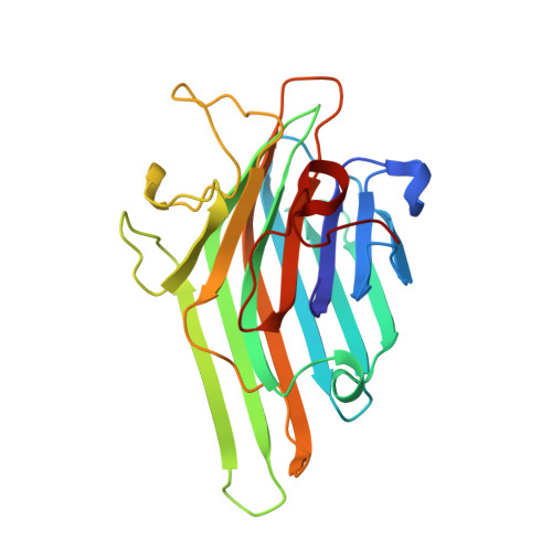
The crystal structure of the complexes of concanavalin A with 4'-nitrophenyl-alpha-D-mannopyranoside and 4'-nitrophenyl-alpha-D-glucopyranoside.

Kanellopoulos, P.N.Pavlou, K.Perrakis, A.Agianian, B.Vorgias, C.E.Mavrommatis, C.Soufi, M.Tucker, P.A.Hamodrakas, S.J.

(1996) J Struct Biol 116: 345-355

  • DOI: https://doi.org/10.1006/jsbi.1996.0052
  • Primary Citation of Related Structures:  
    1VAL, 1VAM

  • PubMed Abstract: 

    Concanavalin A (Con A) is the best-known plant lectin and has important in vitro biological activities arising from its specific saccharide-binding ability. Its exact biological role still remains unknown. The complexes of Con A with 4'-nitrophenyl-alpha-D-mannopyranoside (alpha-PNM) and 4'-nitrophenyl-alpha-D-glucopyranoside (alpha-PNG) have been crystallized in space group P2(1)2(1)2 with cell dimensions a = 135.19 A, b = 155.38 A, c = 71.25 A and a = 134.66 A, b = 155.67 A, and c = 71.42 A, respectively. X-ray diffraction intensities to 2.75 A for the alpha-PNM and to 3.0 A resolution for the alpha-PNG complex have been collected. The structures of the complexes were solved by molecular replacement and refined by simulated annealing methods to crystallographic R-factor values of 0.185/0.186 and free-R-factor values of 0.260/0.274, respectively. In both structures, the asymmetric unit contains four molecules arranged as a tetramer, with approximate 222 symmetry. A saccharide molecule is bound in the sugar-binding site near the surface of each monomer. The nonsugar (aglycon) portion of the compounds used helps to identify the exact orientation of the saccharide in the sugar-binding pocket and is involved in major interactions between tetramers. The hydrogen bonding network in the region of the binding site has been analyzed, and only minor differences with the previously reported Con A-methyl-alpha-D-mannopyranoside complex structure have been observed. Structural differences that may contribute to the slight preference of the lectin for mannosides over glucosides are discussed. Calculations indicate a negative electrostatic surface potential for the saccharide binding site of Con A, which may be important for its biological activity. It is also shown in detail how a particular class of hydrophobic ligands interact with one of the three so-called characteristic hydrophobic sites of the lectins.


  • Organizational Affiliation

    Europeam Molecular Biology Laboratory, Heidelberg, Germany.


Macromolecules
Find similar proteins by:  (by identity cutoff)  |  3D Structure
Entity ID: 1
MoleculeChains Sequence LengthOrganismDetailsImage
CONCANAVALIN A
A, B, C, D
237Canavalia ensiformisMutation(s): 0 
UniProt
Find proteins for P02866 (Canavalia ensiformis)
Explore P02866 
Go to UniProtKB:  P02866
Entity Groups  
Sequence Clusters30% Identity50% Identity70% Identity90% Identity95% Identity100% Identity
UniProt GroupP02866
Sequence Annotations
Expand
  • Reference Sequence
Small Molecules
Ligands 3 Unique
IDChains Name / Formula / InChI Key2D Diagram3D Interactions
PNG
Query on PNG

Download Ideal Coordinates CCD File 
E [auth A],
H [auth B],
K [auth C],
N [auth D]
4-nitrophenyl alpha-D-glucopyranoside
C12 H15 N O8
IFBHRQDFSNCLOZ-ZIQFBCGOSA-N
MN
Query on MN

Download Ideal Coordinates CCD File 
F [auth A],
I [auth B],
L [auth C],
O [auth D]
MANGANESE (II) ION
Mn
WAEMQWOKJMHJLA-UHFFFAOYSA-N
CA
Query on CA

Download Ideal Coordinates CCD File 
G [auth A],
J [auth B],
M [auth C],
P [auth D]
CALCIUM ION
Ca
BHPQYMZQTOCNFJ-UHFFFAOYSA-N
Experimental Data & Validation

Experimental Data

  • Method: X-RAY DIFFRACTION
  • Resolution: 3.00 Å
  • R-Value Free: 0.274 
  • R-Value Work: 0.186 
  • R-Value Observed: 0.186 
  • Space Group: P 21 21 2
Unit Cell:
Length ( Å )Angle ( ˚ )
a = 134.66α = 90
b = 155.67β = 90
c = 71.42γ = 90
Software Package:
Software NamePurpose
X-PLORmodel building
X-PLORrefinement
XDSdata reduction
X-PLORphasing

Structure Validation

View Full Validation Report


Ligand Structure Quality Assessment 


Entry History 

Deposition Data

Revision History  (Full details and data files)

  • Version 1.0: 1997-01-11
    Type: Initial release
  • Version 1.1: 2008-03-24
    Changes: Version format compliance
  • Version 1.2: 2011-07-13
    Changes: Version format compliance
  • Version 1.3: 2020-07-29
    Type: Remediation
    Reason: Carbohydrate remediation
    Changes: Data collection, Database references, Derived calculations, Other, Structure summary
  • Version 1.4: 2024-02-14
    Changes: Data collection, Database references, Structure summary