1V1A

2-KETO-3-DEOXYGLUCONATE KINASE FROM THERMUS THERMOPHILUS WITH BOUND 2-KETO-3-DEOXYGLUCONATE AND ADP


Experimental Data Snapshot

  • Method: X-RAY DIFFRACTION
  • Resolution: 2.10 Å
  • R-Value Free: 0.268 
  • R-Value Work: 0.180 
  • R-Value Observed: 0.180 

wwPDB Validation   3D Report Full Report


Ligand Structure Quality Assessment 


This is version 1.2 of the entry. See complete history


Literature

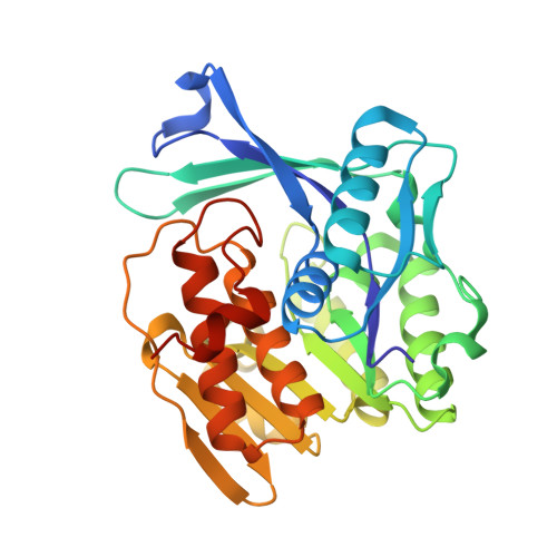
Structure of Thermus thermophilus 2-Keto-3-deoxygluconate kinase: evidence for recognition of an open chain substrate.

Ohshima, N.Inagaki, E.Yasuike, K.Takio, K.Tahirov, T.H.

(2004) J Mol Biol 340: 477-489

  • DOI: https://doi.org/10.1016/j.jmb.2004.04.074
  • Primary Citation of Related Structures:  
    1V19, 1V1A, 1V1B, 1V1S

  • PubMed Abstract: 

    2-Keto-3-deoxygluconate kinase (KDGK) catalyzes the phosphorylation of 2-keto-3-deoxygluconate (KDG) to 2-keto-3-deoxy-6-phosphogluconate (KDGP). The genome sequence of Thermus thermophilus HB8 contains an open reading frame that has a 30% identity to Escherichia coli KDGK. The KDGK activity of T.thermophilus protein (TtKDGK) has been confirmed, and its crystal structure has been determined by the molecular replacement method and refined with two crystal forms to 2.3 angstroms and 3.2 angstroms, respectively. The enzyme is a hexamer organized as a trimer of dimers. Each subunit is composed of two domains, a larger alpha/beta domain and a smaller beta-sheet domain, similar to that of ribokinase and adenosine kinase, members of the PfkB family of carbohydrate kinases. Furthermore, the TtKDGK structure with its KDG and ATP analogue was determined and refined at 2.1 angstroms. The bound KDG was observed predominantly as an open chain structure. The positioning of ligands and the conservation of important catalytic residues suggest that the reaction mechanism is likely to be similar to that of other members of the PfkB family, including ribokinase. In particular, the Asp251 is postulated to have a role in transferring the gamma-phosphate of ATP to the 5'-hydroxyl group of KDG.


  • Organizational Affiliation

    Highthroughput Factory, RIKEN Harima Institute, 1-1-1 Kouto, Mikazuki-cho, Sayo-gun, Hyogo 679-5148, Japan.


Macromolecules
Find similar proteins by:  (by identity cutoff)  |  3D Structure
Entity ID: 1
MoleculeChains Sequence LengthOrganismDetailsImage
2-KETO-3-DEOXYGLUCONATE KINASE
A, B
309Thermus thermophilus HB8Mutation(s): 0 
UniProt
Find proteins for Q53W83 (Thermus thermophilus (strain ATCC 27634 / DSM 579 / HB8))
Explore Q53W83 
Go to UniProtKB:  Q53W83
Entity Groups  
Sequence Clusters30% Identity50% Identity70% Identity90% Identity95% Identity100% Identity
UniProt GroupQ53W83
Sequence Annotations
Expand
  • Reference Sequence
Experimental Data & Validation

Experimental Data

  • Method: X-RAY DIFFRACTION
  • Resolution: 2.10 Å
  • R-Value Free: 0.268 
  • R-Value Work: 0.180 
  • R-Value Observed: 0.180 
  • Space Group: P 63
Unit Cell:
Length ( Å )Angle ( ˚ )
a = 84.321α = 90
b = 84.321β = 90
c = 168.701γ = 120
Software Package:
Software NamePurpose
CNSrefinement
HKL-2000data reduction
HKL-2000data scaling
CNSphasing

Structure Validation

View Full Validation Report


Ligand Structure Quality Assessment 


Entry History 

Deposition Data

Revision History  (Full details and data files)

  • Version 1.0: 2004-04-14
    Type: Initial release
  • Version 1.1: 2015-04-15
    Changes: Data collection, Database references, Derived calculations, Non-polymer description, Other, Refinement description, Version format compliance
  • Version 1.2: 2019-02-27
    Changes: Data collection, Database references, Experimental preparation, Other