1UZ3

Crystal structure of novel protein EMSY


Experimental Data Snapshot

  • Method: X-RAY DIFFRACTION
  • Resolution: 1.10 Å
  • R-Value Free: 0.199 
  • R-Value Observed: 0.151 

wwPDB Validation   3D Report Full Report


This is version 1.3 of the entry. See complete history


Literature

Crystal Structure of the Ent Domain of Human Emsy.

Chavali, G.B.Ekblad, C.M.Basu, B.P.Brissett, N.C.Veprintsev, D.Hughes-Davies, L.Kouzarides, T.Itzhaki, L.S.Doherty, A.J.

(2005) J Mol Biol 350: 964

  • DOI: https://doi.org/10.1016/j.jmb.2005.05.047
  • Primary Citation of Related Structures:  
    1UZ3

  • PubMed Abstract: 

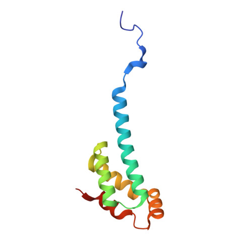
    EMSY is a recently discovered gene encoding a BRCA2-associated protein and is amplified in some sporadic breast and ovarian cancers. The EMSY sequence contains no known domain except for a conserved approximately 100 residue segment at the N terminus. This so-called ENT domain is unique in the human genome, although multiple copies are found in Arabidopsis proteins containing members of the Royal family of chromatin remodelling domains. Here, we report the crystal structure of the ENT domain of EMSY, consisting of a unique arrangement of five alpha-helices that fold into a helical bundle arrangement. The fold shares regions of structural homology with the DNA-binding domain of homeodomain proteins. The ENT domain forms a homodimer via the anti-parallel packing of the extended N-terminal alpha-helix of each molecule. It is stabilized mainly by hydrophobic residues at the dimer interface and has a dissociation constant in the low micromolar range. The dimerisation of EMSY mediated by the ENT domain could provide flexibility for it to bind two or more different substrates simultaneously.


  • Organizational Affiliation

    Department of Haematology, Cambridge Institute for Medical Research, University of Cambridge, Hills Rd, Cambridge CB2 2XY, UK.


Macromolecules
Find similar proteins by:  (by identity cutoff)  |  3D Structure
Entity ID: 1
MoleculeChains Sequence LengthOrganismDetailsImage
EMSY PROTEIN
A, B
102Homo sapiensMutation(s): 0 
UniProt & NIH Common Fund Data Resources
Find proteins for Q7Z589 (Homo sapiens)
Explore Q7Z589 
Go to UniProtKB:  Q7Z589
PHAROS:  Q7Z589
GTEx:  ENSG00000158636 
Entity Groups  
Sequence Clusters30% Identity50% Identity70% Identity90% Identity95% Identity100% Identity
UniProt GroupQ7Z589
Sequence Annotations
Expand
  • Reference Sequence
Experimental Data & Validation

Experimental Data

  • Method: X-RAY DIFFRACTION
  • Resolution: 1.10 Å
  • R-Value Free: 0.199 
  • R-Value Observed: 0.151 
  • Space Group: P 1 21 1
Unit Cell:
Length ( Å )Angle ( ˚ )
a = 31.643α = 90
b = 55.538β = 92.53
c = 51.278γ = 90
Software Package:
Software NamePurpose
SHELXL-97refinement
MOSFLMdata reduction
SCALAdata scaling
ACORNphasing

Structure Validation

View Full Validation Report


Entry History 

Deposition Data

Revision History  (Full details and data files)

  • Version 1.0: 2005-10-13
    Type: Initial release
  • Version 1.1: 2011-05-07
    Changes: Version format compliance
  • Version 1.2: 2011-07-13
    Changes: Version format compliance
  • Version 1.3: 2019-05-22
    Changes: Data collection, Other, Refinement description