1UWX

P1.2 serosubtype antigen derived from N. meningitidis PorA in complex with Fab fragment


Experimental Data Snapshot

  • Method: X-RAY DIFFRACTION
  • Resolution: 2.20 Å
  • R-Value Free: 0.265 
  • R-Value Work: 0.223 
  • R-Value Observed: 0.225 

wwPDB Validation   3D Report Full Report


This is version 1.2 of the entry. See complete history


Literature

Structural Variation and Immune Recognition of the P1.2 Subtype Meningococcal Antigen.

Tzitzilonis, C.Prince, S.M.Collins, R.F.Achtman, M.Feavers, I.M.Maiden, M.C.J.Derrick, J.P.

(2005) Proteins 62: 947

  • DOI: https://doi.org/10.1002/prot.20800
  • Primary Citation of Related Structures:  
    1UWX

  • PubMed Abstract: 

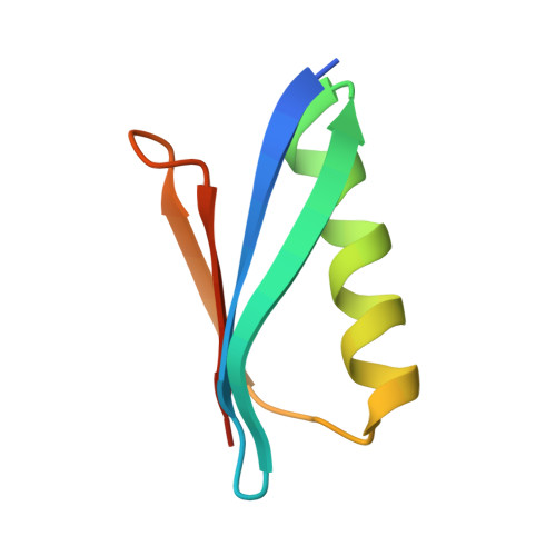
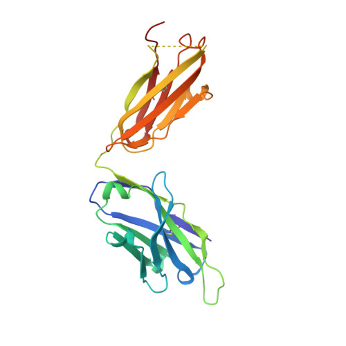
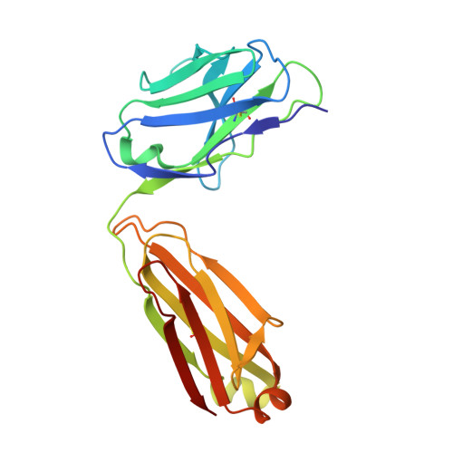
    Neisseria meningitidis is a globally important cause of bacterial meningitis and septicemia. No comprehensive antimeningococcal vaccine is available, largely as a consequence of the high sequence diversity of those surface proteins that could function as components of a vaccine. One such component is the protein PorA, a major surface porin of this Gram-negative organism that has been used in a number of experimental and licensed vaccines. Here we describe a series of experiments designed to investigate the consequences for antibody recognition of sequence diversity within a PorA antigen. The binding of a 14-residue peptide, corresponding to the P1.2 subtype antigen, to the MN16C13F4 monoclonal antibody was sensitive to mutation of five out of the six residues within the epitope sequence. The crystal structure of the antibody Fab fragment, determined in complex with the peptide antigen, shows a remarkably hydrophobic binding site and interactions between the antigen and antibody are dominated by apolar residues. Nine intrachain hydrogen bonds are formed within the antigen which maintain the beta-hairpin conformation of the peptide. These hydrogen bonds involve residues that are highly conserved amongst different P1.2 sequence variants, suggesting that some positions may be conserved for structural reasons in these highly polymorphic regions. The sensitivity of antibody recognition of the antigen towards mutation provides a structural explanation for the widespread sequence variation seen in different PorA sequences in this region. Single point mutations are sufficient to remove binding capability, providing a rationale for the manner in which different meningococcal PorA escape variants arise.


  • Organizational Affiliation

    Faculty of Life Sciences, The University of Manchester, Manchester, United Kingdom.


Macromolecules
Find similar proteins by:  (by identity cutoff)  |  3D Structure
Entity ID: 1
MoleculeChains Sequence LengthOrganismDetailsImage
PROTEIN G-PRIME
A, B
63Streptococcus sp.Mutation(s): 1 
UniProt
Find proteins for P19909 (Streptococcus sp. group G)
Explore P19909 
Go to UniProtKB:  P19909
Entity Groups  
Sequence Clusters30% Identity50% Identity70% Identity90% Identity95% Identity100% Identity
UniProt GroupP19909
Sequence Annotations
Expand
  • Reference Sequence
Find similar proteins by:  (by identity cutoff)  |  3D Structure
Entity ID: 2
MoleculeChains Sequence LengthOrganismDetailsImage
ANTIBODYC [auth H],
F [auth M]
225Mus musculusMutation(s): 0 
Entity Groups  
Sequence Clusters30% Identity50% Identity70% Identity90% Identity95% Identity100% Identity
Sequence Annotations
Expand
  • Reference Sequence
Find similar proteins by:  (by identity cutoff)  |  3D Structure
Entity ID: 3
MoleculeChains Sequence LengthOrganismDetailsImage
ANTIBODYD [auth K],
E [auth L]
213Mus musculusMutation(s): 0 
UniProt
Find proteins for P01837 (Mus musculus)
Explore P01837 
Go to UniProtKB:  P01837
Entity Groups  
Sequence Clusters30% Identity50% Identity70% Identity90% Identity95% Identity100% Identity
UniProt GroupP01837
Sequence Annotations
Expand
  • Reference Sequence

Find similar proteins by:  Sequence   |   3D Structure  

Entity ID: 4
MoleculeChains Sequence LengthOrganismDetailsImage
CLASS 1 OUTER MEMBRANE PROTEIN VARIABLE REGION 2G [auth P],
H [auth Q]
13Neisseria meningitidisMutation(s): 0 
UniProt
Find proteins for Q9JPT2 (Neisseria meningitidis)
Explore Q9JPT2 
Go to UniProtKB:  Q9JPT2
Entity Groups  
Sequence Clusters30% Identity50% Identity70% Identity90% Identity95% Identity100% Identity
UniProt GroupQ9JPT2
Sequence Annotations
Expand
  • Reference Sequence
Experimental Data & Validation

Experimental Data

  • Method: X-RAY DIFFRACTION
  • Resolution: 2.20 Å
  • R-Value Free: 0.265 
  • R-Value Work: 0.223 
  • R-Value Observed: 0.225 
  • Space Group: P 21 21 21
Unit Cell:
Length ( Å )Angle ( ˚ )
a = 81.426α = 90
b = 110.568β = 90
c = 138.734γ = 90
Software Package:
Software NamePurpose
REFMACrefinement
MOSFLMdata reduction
SCALAdata scaling
AMoREphasing

Structure Validation

View Full Validation Report


Entry History 

Deposition Data

Revision History  (Full details and data files)

  • Version 1.0: 2005-06-15
    Type: Initial release
  • Version 1.1: 2011-07-13
    Changes: Refinement description, Version format compliance
  • Version 1.2: 2023-12-13
    Changes: Data collection, Database references, Refinement description