Structural difference between group I and group II cobra cardiotoxins: X-ray, NMR, and CD analysis of the effect of cis-proline conformation on three-fingered toxins.
Chen, T.S., Chung, F.Y., Tjong, S.C., Goh, K.S., Huang, W.N., Chien, K.Y., Wu, P.L., Lin, H.C., Chen, C.J., Wu, W.G.(2005) Biochemistry 44: 7414-7426
- PubMed: 15895985
- DOI: https://doi.org/10.1021/bi050172e
- Primary Citation of Related Structures:
1UG4 - PubMed Abstract:
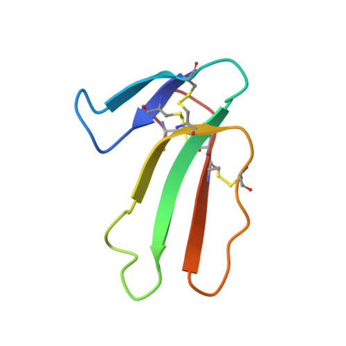
Natural homologues of cobra cardiotoxins (CTXs) were classified into two structural subclasses of group I and II based on the amino acid sequence and circular dichroism analysis, but the exact differences in their three-dimensional structures and biological significance remain elusive. We show by circular dichroism, NMR spectroscopic, and X-ray crystallographic analyses of a newly purified group I CTX A6 from eastern Taiwan cobra (Naja atra) venoms that its loop I conformation adopts a type VIa turn with a cis peptide bond located between two proline residues of PPxY. A similar "banana-twisted" conformation can be observed in other group I CTXs and also in cyclolinopeptide A and its analogues. By binding to the membrane environment, group I CTX undergoes a conformational change to adopt a more extended hydrophobic domain with beta-sheet twisting closer to the one adopted by group II CTX. This result resolves a discrepancy in the CTX structural difference reported previously between solution as well as crystal state and shows that, in addition to the hydrophobicity, the exact loop I conformation also plays an important role in CTX-membrane interaction. Potential protein targets of group I CTXs after cell internalization are also discussed on the basis of the determined loop I conformation.
Organizational Affiliation:
Institute of Bioinformatics and Structural Biology, National Tsing Hua University, Hsinchu 300, Taiwan.