1UEI

Crystal structure of human uridine-cytidine kinase 2 complexed with a feedback-inhibitor, UTP


Experimental Data Snapshot

  • Method: X-RAY DIFFRACTION
  • Resolution: 2.60 Å
  • R-Value Free: 0.245 
  • R-Value Work: 0.204 
  • R-Value Observed: 0.204 

wwPDB Validation   3D Report Full Report


Ligand Structure Quality Assessment 


This is version 1.3 of the entry. See complete history


Literature

Structural basis for the specificity, catalysis, and regulation of human uridine-cytidine kinase

Suzuki, N.N.Koizumi, K.Fukushima, M.Matsuda, A.Inagaki, F.

(2004) Structure 12: 751-764

  • DOI: https://doi.org/10.1016/j.str.2004.02.038
  • Primary Citation of Related Structures:  
    1UDW, 1UEI, 1UEJ, 1UFQ, 1UJ2

  • PubMed Abstract: 

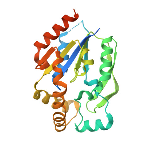
    Uridine-cytidine kinase (UCK) catalyzes the phosphorylation of uridine and cytidine and activates pharmacological ribonucleoside analogs. Here we present the crystal structures of human UCK alone and in complexes with a substrate, cytidine, a feedback inhibitor, CTP or UTP, and with phosphorylation products, CMP and ADP, respectively. Free UCK takes an alpha/beta mononucleotide binding fold and exists as a homotetramer with 222 symmetry. Upon inhibitor binding, one loop region was loosened, causing the UCK tetramer to be distorted. Upon cytidine binding, a large induced fit was observed at the uridine/cytidine binding site, which endows UCK with a strict specificity for pyrimidine ribonucleosides. The first UCK structure provided the structural basis for the specificity, catalysis, and regulation of human uridine-cytidine kinase, which give clues for the design of novel antitumor and antiviral ribonucleoside analogs that inhibit RNA synthesis.


  • Organizational Affiliation

    Department of Structural Biology, Graduate School of Pharmaceutical Sciences, Hokkaido University, N12 W6 Kita-ku, Sapporo 060-0812, Japan.


Macromolecules
Find similar proteins by:  (by identity cutoff)  |  3D Structure
Entity ID: 1
MoleculeChains Sequence LengthOrganismDetailsImage
Uridine-cytidine kinase 2
A, B
252Homo sapiensMutation(s): 0 
EC: 2.7.1.48
UniProt & NIH Common Fund Data Resources
Find proteins for Q9BZX2 (Homo sapiens)
Explore Q9BZX2 
Go to UniProtKB:  Q9BZX2
PHAROS:  Q9BZX2
GTEx:  ENSG00000143179 
Entity Groups  
Sequence Clusters30% Identity50% Identity70% Identity90% Identity95% Identity100% Identity
UniProt GroupQ9BZX2
Sequence Annotations
Expand
  • Reference Sequence
Small Molecules
Experimental Data & Validation

Experimental Data

  • Method: X-RAY DIFFRACTION
  • Resolution: 2.60 Å
  • R-Value Free: 0.245 
  • R-Value Work: 0.204 
  • R-Value Observed: 0.204 
  • Space Group: F 2 2 2
Unit Cell:
Length ( Å )Angle ( ˚ )
a = 131.529α = 90
b = 247.748β = 90
c = 91.673γ = 90
Software Package:
Software NamePurpose
CNSrefinement
CrystalCleardata reduction
CrystalCleardata scaling
CNSphasing

Structure Validation

View Full Validation Report


Ligand Structure Quality Assessment 


Entry History 

Deposition Data

Revision History  (Full details and data files)

  • Version 1.0: 2004-05-04
    Type: Initial release
  • Version 1.1: 2008-04-27
    Changes: Version format compliance
  • Version 1.2: 2011-07-13
    Changes: Version format compliance
  • Version 1.3: 2023-10-25
    Changes: Data collection, Database references, Derived calculations, Refinement description