1U9R

Crystal Structure of Staphylococcal Nuclease mutant V66E/P117G/H124L/S128A at Room Temperature


Experimental Data Snapshot

  • Method: X-RAY DIFFRACTION
  • Resolution: 2.10 Å
  • R-Value Free: 0.217 
  • R-Value Work: 0.193 
  • R-Value Observed: 0.193 

wwPDB Validation   3D Report Full Report


This is version 1.6 of the entry. See complete history


Literature

Stabilization of internal charges in a protein: water penetration or conformational change?

Denisov, V.P.Schlessman, J.L.Garcia-Moreno, B.E.Halle, B.

(2004) Biophys J 87: 3982-3994

  • DOI: https://doi.org/10.1529/biophysj.104.048454
  • Primary Citation of Related Structures:  
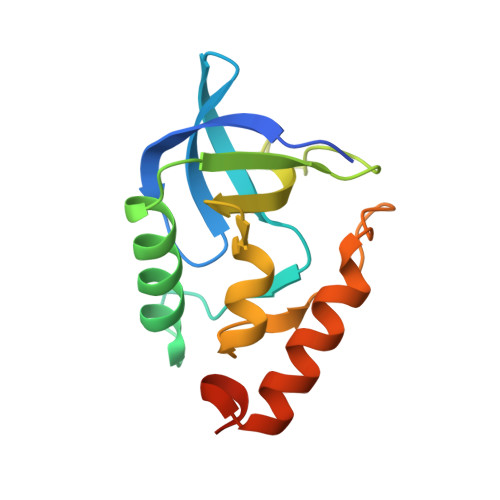
    1U9R

  • PubMed Abstract: 

    The ionizable amino acid side chains of proteins are usually located at the surface. However, in some proteins an ionizable group is embedded in an apolar internal region. Such buried ionizable groups destabilize the protein and may trigger conformational changes in response to pH variations. Because of the prohibitive energetic cost of transferring a charged group from water to an apolar medium, other stabilizing factors must be invoked, such as ionization-induced water penetration or structural changes. To examine the role of water penetration, we have measured the 17O and 2H magnetic relaxation dispersions (MRD) for the V66E and V66K mutants of staphylococcal nuclease, where glutamic acid and lysine residues are buried in predominantly apolar environments. At neutral pH, where these residues are uncharged, we find no evidence of buried water molecules near the mutation site. This contrasts with a previous cryogenic crystal structure of the V66E mutant, but is consistent with the room-temperature crystal structure reported here. MRD measurements at different pH values show that ionization of Glu-66 or Lys-66 is not accompanied by penetration of long-lived water molecules. On the other hand, the MRD data are consistent with a local conformational change in response to ionization of the internal residues.


  • Organizational Affiliation

    Department of Biophysical Chemistry, Lund University, Lund, Sweden.


Macromolecules
Find similar proteins by:  (by identity cutoff)  |  3D Structure
Entity ID: 1
MoleculeChains Sequence LengthOrganismDetailsImage
Thermonuclease149Staphylococcus aureusMutation(s): 4 
Gene Names: nuc
EC: 3.1.31.1
UniProt
Find proteins for P00644 (Staphylococcus aureus)
Explore P00644 
Go to UniProtKB:  P00644
Entity Groups  
Sequence Clusters30% Identity50% Identity70% Identity90% Identity95% Identity100% Identity
UniProt GroupP00644
Sequence Annotations
Expand
  • Reference Sequence
Experimental Data & Validation

Experimental Data

  • Method: X-RAY DIFFRACTION
  • Resolution: 2.10 Å
  • R-Value Free: 0.217 
  • R-Value Work: 0.193 
  • R-Value Observed: 0.193 
  • Space Group: P 41
Unit Cell:
Length ( Å )Angle ( ˚ )
a = 48.794α = 90
b = 48.794β = 90
c = 63.788γ = 90
Software Package:
Software NamePurpose
CNSrefinement
DENZOdata reduction
SCALEPACKdata scaling
CNSphasing

Structure Validation

View Full Validation Report


Entry History 

Deposition Data

Revision History  (Full details and data files)

  • Version 1.0: 2004-12-21
    Type: Initial release
  • Version 1.1: 2008-04-30
    Changes: Version format compliance
  • Version 1.2: 2011-07-13
    Changes: Version format compliance
  • Version 1.3: 2017-10-11
    Changes: Refinement description
  • Version 1.4: 2021-10-20
    Changes: Database references
  • Version 1.5: 2024-02-14
    Changes: Data collection
  • Version 1.6: 2024-04-03
    Changes: Refinement description