Solution structure of the fibrin binding finger domain of tissue-type plasminogen activator determined by 1H nuclear magnetic resonance.
Downing, A.K., Driscoll, P.C., Harvey, T.S., Dudgeon, T.J., Smith, B.O., Baron, M., Campbell, I.D.(1992) J Mol Biol 225: 821-833
- PubMed: 1602484
- DOI: https://doi.org/10.1016/0022-2836(92)90403-7
- Primary Citation of Related Structures:
1TPM, 1TPN - PubMed Abstract:
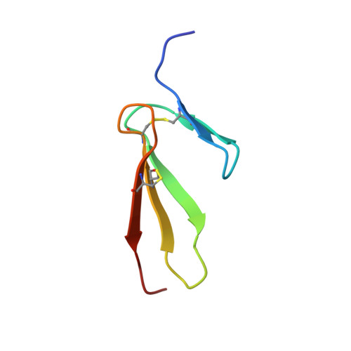
The amino acid sequence of the first domain of tissue-type plasminogen activator (t-PA) includes eight residues that are highly conserved in the type 1 finger domains found in human fibronectin. A construct comprising 50 residues from this finger domain of t-PA has been expressed and its solution structure has been determined by two-dimensional nuclear magnetic resonance spectroscopy. A total of 782 experimental restraints consisting of 723 interproton distances derived from nuclear Overhauser effect measurements, 43 torsion angles, and 16 hydrogen bond restraints were used as the input for dynamical simulated annealing structure calculations. Twenty-eight structures were obtained that satisfied the experimental data with no single distance violation greater than 0.3 A. The average atomic root-mean-square distribution for the backbone atoms of the final structures was 0.41 (+/- 0.13) A for the well defined part of the structure (residues 4 to 47). The overall fold of the t-PA finger domain shows a striking similarity to that of the seventh type 1 repeat of human fibronectin with the side-chains of conserved residues lying in similar conformations. One significant difference between the two molecules is that hydrophobic residues cover the exposed surface of the principal beta-sheet region in the t-PA finger domain. It is suggested that one face of this region may interact with parts of the complete t-PA protein.
Organizational Affiliation:
Department of Biochemistry, University of Oxford, U.K.