1TPE

COMPARISON OF THE STRUCTURES AND THE CRYSTAL CONTACTS OF TRYPANOSOMAL TRIOSEPHOSPHATE ISOMERASE IN FOUR DIFFERENT CRYSTAL FORMS


Experimental Data Snapshot

  • Method: X-RAY DIFFRACTION
  • Resolution: 2.10 Å
  • R-Value Work: 0.155 
  • R-Value Observed: 0.155 

wwPDB Validation   3D Report Full Report


This is version 1.5 of the entry. See complete history


Literature

Comparison of the structures and the crystal contacts of trypanosomal triosephosphate isomerase in four different crystal forms.

Kishan, K.V.Zeelen, J.P.Noble, M.E.Borchert, T.V.Wierenga, R.K.

(1994) Protein Sci 3: 779-787

  • DOI: https://doi.org/10.1002/pro.5560030507
  • Primary Citation of Related Structures:  
    1TPE, 1TPF

  • PubMed Abstract: 

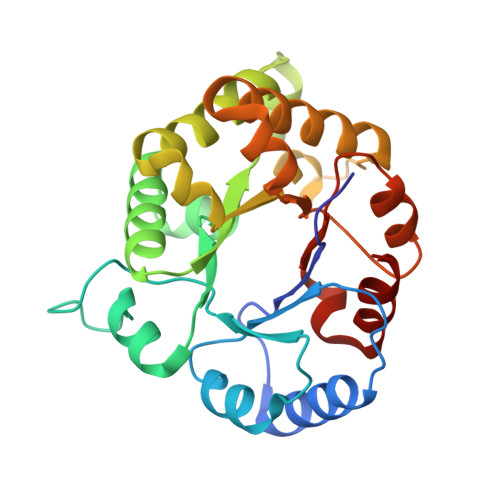
    Triosephosphate isomerase (TIM) is a dimeric enzyme consisting of 2 identical subunits. Trypanosomal TIM can be crystallized in 4 different spacegroups: P2(1)2(1)2(1), C2(big cell), C2(small cell), and P1. The P1 crystal form only grows in the presence of 1.4 M DMSO; there are 2 DMSO binding sites per subunit. The structures have been refined at a resolution of 1.83 A, 2.10 A, 2.13 A, and 1.80 A, respectively. In the 4 different spacegroups the TIM subunit can be observed in the context of 7 different crystallographic environments. In the C2 cells, the dimer 2-fold axis coincides with a crystallographic 2-fold axis. The similarities and differences of the 7 subunits are discussed. In 6 subunits the flexible loop (loop 6) is open, whereas in the P2(1)2(1)2(1) cell, the flexible loop of subunit 2 is in an almost closed conformation. The crystal contacts in the 4 different crystal forms are predominantly generated by polar residues in loops. A statistical analysis of the residues involved in crystal contacts shows that, in particular, serines are frequently involved in these interactions; 19% of the exposed serines are involved in crystal contacts.


  • Organizational Affiliation

    European Molecular Biology Laboratory, Heidelberg, Germany.


Macromolecules
Find similar proteins by:  (by identity cutoff)  |  3D Structure
Entity ID: 1
MoleculeChains Sequence LengthOrganismDetailsImage
TRIOSEPHOSPHATE ISOMERASE250Trypanosoma brucei bruceiMutation(s): 0 
EC: 5.3.1.1
UniProt
Find proteins for P04789 (Trypanosoma brucei brucei)
Explore P04789 
Go to UniProtKB:  P04789
Entity Groups  
Sequence Clusters30% Identity50% Identity70% Identity90% Identity95% Identity100% Identity
UniProt GroupP04789
Sequence Annotations
Expand
  • Reference Sequence
Experimental Data & Validation

Experimental Data

  • Method: X-RAY DIFFRACTION
  • Resolution: 2.10 Å
  • R-Value Work: 0.155 
  • R-Value Observed: 0.155 
  • Space Group: C 1 2 1
Unit Cell:
Length ( Å )Angle ( ˚ )
a = 95.4α = 90
b = 48.2β = 120
c = 67.5γ = 90
Software Package:
Software NamePurpose
X-PLORmodel building
TNTrefinement
X-PLORrefinement
X-PLORphasing

Structure Validation

View Full Validation Report


Entry History 

Deposition Data

Revision History  (Full details and data files)

  • Version 1.0: 1994-05-31
    Type: Initial release
  • Version 1.1: 2008-03-24
    Changes: Version format compliance
  • Version 1.2: 2011-07-13
    Changes: Derived calculations, Version format compliance
  • Version 1.3: 2019-07-17
    Changes: Data collection, Other, Refinement description
  • Version 1.4: 2019-08-14
    Changes: Data collection, Refinement description
  • Version 1.5: 2024-02-14
    Changes: Data collection, Database references