1TLV

Structure of the native and inactive LicT PRD from B. subtilis


Experimental Data Snapshot

  • Method: X-RAY DIFFRACTION
  • Resolution: 1.95 Å
  • R-Value Free: 0.292 
  • R-Value Work: 0.269 
  • R-Value Observed: 0.269 

wwPDB Validation   3D Report Full Report


This is version 1.2 of the entry. See complete history

Re-refinement Note

A newer entry is available that reflects an alternative modeling of the original data: 6TWR


Literature

Activation of the LicT Transcriptional Antiterminator Involves a Domain Swing/Lock Mechanism Provoking Massive Structural Changes

Graille, M.Zhou, C.-Z.Receveur-Brechot, V.Collinet, B.Declerck, N.van Tilbeurgh, H.

(2005) J Biol Chem 280: 14780-14789

  • DOI: https://doi.org/10.1074/jbc.M414642200
  • Primary Citation of Related Structures:  
    1TLV

  • PubMed Abstract: 

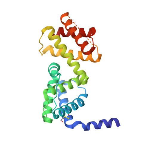
    The transcriptional antiterminator protein LicT regulates the expression of Bacillus subtilis operons involved in beta-glucoside metabolism. It consists of an N-terminal RNA-binding domain (co-antiterminator (CAT)) and two phosphorylatable phosphotransferase system regulation domains (PRD1 and PRD2). In the activated state, each PRD forms a dimeric unit with the phosphorylation sites totally buried at the dimer interface. Here we present the 1.95 A resolution structure of the inactive LicT PRDs as well as the molecular solution structure of the full-length protein deduced from small angle x-ray scattering. Comparison of native (inactive) and mutant (constitutively active) PRD crystal structures shows massive tertiary and quaternary rearrangements of the entire regulatory domain. In the inactive state, a wide swing movement of PRD2 results in dimer opening and brings the phosphorylation sites to the protein surface. This movement is accompanied by additional structural rearrangements of both the PRD1-PRD1 ' interface and the CAT-PRD1 linker. Small angle x-ray scattering experiments indicate that the amplitude of the PRD2 swing might even be wider in solution than in the crystals. Our results suggest that PRD2 is highly mobile in the native protein, whereas it is locked upon activation by phosphorylation.


  • Organizational Affiliation

    Laboratoire d'Enzymologie et Biochimie Structurales, CNRS-UPR9063, Gif sur Yvette, France.


Macromolecules
Find similar proteins by:  (by identity cutoff)  |  3D Structure
Entity ID: 1
MoleculeChains Sequence LengthOrganismDetailsImage
Transcription antiterminator licT221Bacillus subtilisMutation(s): 1 
Gene Names: LICTN15ABSU39080
UniProt
Find proteins for P39805 (Bacillus subtilis (strain 168))
Explore P39805 
Go to UniProtKB:  P39805
Entity Groups  
Sequence Clusters30% Identity50% Identity70% Identity90% Identity95% Identity100% Identity
UniProt GroupP39805
Sequence Annotations
Expand
  • Reference Sequence
Small Molecules
Modified Residues  1 Unique
IDChains TypeFormula2D DiagramParent
CME
Query on CME
A
L-PEPTIDE LINKINGC5 H11 N O3 S2CYS
Experimental Data & Validation

Experimental Data

  • Method: X-RAY DIFFRACTION
  • Resolution: 1.95 Å
  • R-Value Free: 0.292 
  • R-Value Work: 0.269 
  • R-Value Observed: 0.269 
  • Space Group: P 32 2 1
Unit Cell:
Length ( Å )Angle ( ˚ )
a = 48.734α = 90
b = 48.734β = 90
c = 162.472γ = 120
Software Package:
Software NamePurpose
CNSrefinement
DENZOdata reduction
SCALEPACKdata scaling
SOLVEphasing

Structure Validation

View Full Validation Report


Entry History 

Deposition Data

Revision History  (Full details and data files)

  • Version 1.0: 2005-02-08
    Type: Initial release
  • Version 1.1: 2008-04-30
    Changes: Version format compliance
  • Version 1.2: 2011-07-13
    Changes: Derived calculations, Version format compliance