1T92

Crystal structure of N-terminal truncated pseudopilin PulG


Experimental Data Snapshot

  • Method: X-RAY DIFFRACTION
  • Resolution: 1.60 Å
  • R-Value Free: 0.198 
  • R-Value Work: 0.167 
  • R-Value Observed: 0.169 

wwPDB Validation   3D Report Full Report


This is version 1.4 of the entry. See complete history


Literature

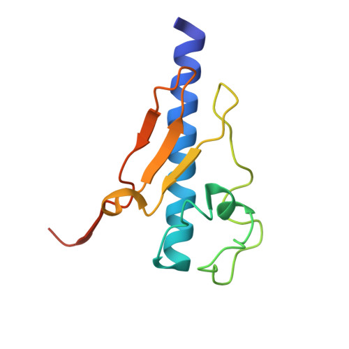
Structure and assembly of the pseudopilin PulG.

Koehler, R.Schaefer, K.Mueller, S.Vignon, G.Diederichs, K.Philippsen, A.Ringler, P.Pugsley, A.P.Engel, A.Welte, W.

(2004) Mol Microbiol 54: 647-664

  • DOI: https://doi.org/10.1111/j.1365-2958.2004.04307.x
  • Primary Citation of Related Structures:  
    1T92

  • PubMed Abstract: 

    The pseudopilin PulG is one of several essential components of the type II pullulanase secretion machinery (the Pul secreton) of the Gram-negative bacterium Klebsiella oxytoca. The sequence of the N-terminal 25 amino acids of the PulG precursor is hydrophobic and very similar to the corresponding region of type IV pilins. The structure of a truncated PulG (lacking the homologous region), as determined by X-ray crystallography, was found to include part of the long N-terminal alpha-helix and the four internal anti-parallel beta-strands that characterize type IV pilins, but PulG lacks the highly variable loop region with a disulphide bond that is found in the latter. When overproduced, PulG forms flexible pili whose structural features, as visualized by electron microscopy, are similar to those of bacterial type IV pili. The average helical repeat comprises 17 PulG subunits and four helical turns. Electron microscopy and molecular modelling show that PulG probably assembles into left-handed helical pili with the long N-terminal alpha-helix tightly packed in the centre of the pilus. As in the type IV pilins, the hydrophobic N-terminal part of the PulG alpha-helix is necessary for its assembly. Subtle sequence variations within this highly conserved segment seem to determine whether or not a type IV pilin can be assembled into pili by the Pul secreton.


  • Organizational Affiliation

    Molecular Genetics Unit, Institut Pasteur, 25, rue du Dr Roux, 75724 Paris Cedex 15, France.


Macromolecules
Find similar proteins by:  (by identity cutoff)  |  3D Structure
Entity ID: 1
MoleculeChains Sequence LengthOrganismDetailsImage
General secretion pathway protein G
A, B
116Klebsiella pneumoniaeMutation(s): 0 
Gene Names: PULG
UniProt
Find proteins for P15746 (Klebsiella pneumoniae)
Explore P15746 
Go to UniProtKB:  P15746
Entity Groups  
Sequence Clusters30% Identity50% Identity70% Identity90% Identity95% Identity100% Identity
UniProt GroupP15746
Sequence Annotations
Expand
  • Reference Sequence
Small Molecules
Ligands 1 Unique
IDChains Name / Formula / InChI Key2D Diagram3D Interactions
ZN
Query on ZN

Download Ideal Coordinates CCD File 
C [auth A]ZINC ION
Zn
PTFCDOFLOPIGGS-UHFFFAOYSA-N
Experimental Data & Validation

Experimental Data

  • Method: X-RAY DIFFRACTION
  • Resolution: 1.60 Å
  • R-Value Free: 0.198 
  • R-Value Work: 0.167 
  • R-Value Observed: 0.169 
  • Space Group: P 65 2 2
Unit Cell:
Length ( Å )Angle ( ˚ )
a = 85.395α = 90
b = 85.395β = 90
c = 145.7205γ = 120
Software Package:
Software NamePurpose
REFMACrefinement
XDSdata reduction
XSCALEdata scaling
SHELXDphasing
SHELXEmodel building

Structure Validation

View Full Validation Report


Entry History 

Deposition Data

Revision History  (Full details and data files)

  • Version 1.0: 2004-10-05
    Type: Initial release
  • Version 1.1: 2008-04-30
    Changes: Version format compliance
  • Version 1.2: 2011-07-13
    Changes: Version format compliance
  • Version 1.3: 2018-01-31
    Changes: Experimental preparation
  • Version 1.4: 2024-02-14
    Changes: Data collection, Database references, Derived calculations, Refinement description