Crystallographic and NMR Investigation of Cobalt-Substituted Amicyanin.
Carrell, C.J., Wang, X., Jones, L., Jarrett, W.L., Davidson, V.L., Mathews, F.S.(2004) Biochemistry 43: 9381-9389
- PubMed: 15260481
- DOI: https://doi.org/10.1021/bi049635r
- Primary Citation of Related Structures:
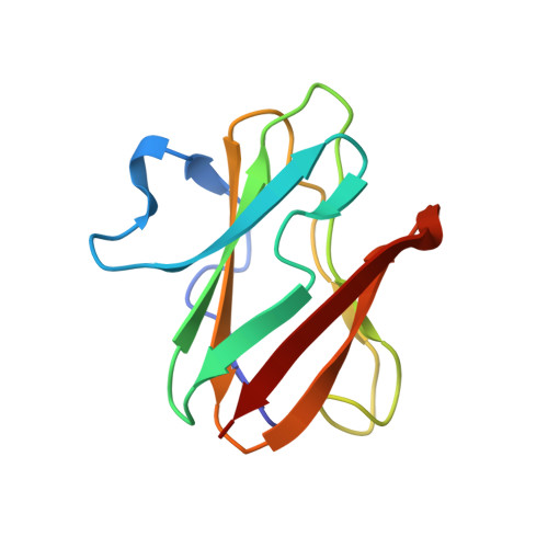
1T5K - PubMed Abstract:
Cobalt(II) amicyanin was prepared by replacing the copper of the type I copper protein amicyanin from Paracoccus denitrificans with cobalt. The structure of the protein and the metal center have been characterized by X-ray crystallography and paramagnetic NMR spectroscopy. The crystal structure indicates that Met98, which provides an axial sulfur ligand in native amicyanin, is no longer bound to the metal in cobalt(II) amicyanin and that a water molecule is recruited from solvent to form the fourth metal ligand. This results in a tetrahedral coordination geometry for the cobalt ion. NMR studies in solution also indicate that the side chain of the methionine residue interacts less strongly with the metal in P. denitrificans amicyanin than in Paracoccus versutus amicyanin. The cobalt(II) amicyanin crystal structure is different from that of cobalt-substituted azurin in which the carbonyl of a glycine residue provides this equivalent ligand. In cobalt(II) amicyanin that residue is a proline, for which the oxygen is structurally inaccessible, so that the water occupies the position held by the glycine carbonyl in cobalt(II) azurin. Such a metal coordination involving water has not previously been reported for a native or metal-substituted type I copper protein.
Organizational Affiliation:
Department of Biochemistry and Molecular Biophysics, Washington University School of Medicine, St. Louis, Missouri 63110, USA.