1SL3

crystal structue of Thrombin in complex with a potent P1 heterocycle-Aryl based inhibitor


Experimental Data Snapshot

  • Method: X-RAY DIFFRACTION
  • Resolution: 1.81 Å
  • R-Value Free: 0.240 
  • R-Value Work: 0.210 
  • R-Value Observed: 0.211 

wwPDB Validation   3D Report Full Report


This is version 1.3 of the entry. See complete history


Literature

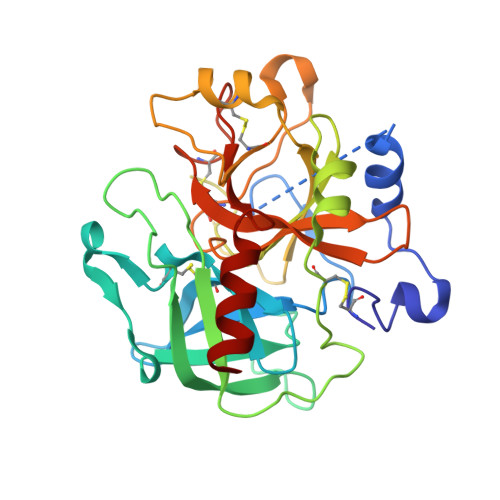
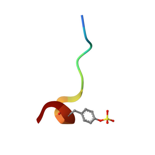
Discovery and evaluation of potent P1 aryl heterocycle-based thrombin inhibitors

Young, M.B.Barrow, J.C.Glass, K.L.Lundell, G.F.Newton, C.L.Pellicore, J.M.Rittle, K.E.Selnick, H.G.Stauffer, K.J.Vacca, J.P.Williams, P.D.Bohn, D.Clayton, F.C.Cook, J.J.Krueger, J.A.Kuo, L.C.Lewis, S.D.Lucas, B.J.McMasters, D.R.Miller-Stein, C.Pietrak, B.L.

(2004) J Med Chem 47: 2995-3008

  • DOI: https://doi.org/10.1021/jm030303e
  • Primary Citation of Related Structures:  
    1SL3

  • PubMed Abstract: 

    In an effort to discover potent, clinically useful thrombin inhibitors, a rapid analogue synthetic approach was used to explore the P(1) region. Various benzylamines were coupled to a pyridine/pyrazinone P(2)-P(3) template. One compound with an o-thiadiazole benzylic substitution was found to have a thrombin K(i) of 0.84 nM. A study of ortho-substituted five-membered-ring heterocycles was undertaken and subsequently demonstrated that the o-triazole and tetrazole rings were optimal. Combination of these potent P(1) aryl heterocycles with a variety of P(2)-P(3) groups produced a compound with an extraordinary thrombin inhibitory activity of 1.4 pM. It is hoped that this potency enhancement in P(1) will allow for more diversification in the P(2)-P(3) region to ultimately address additional pharmacological concerns.


  • Organizational Affiliation

    Medicinal Chemistry, Merck Research Laboratories, Merck and Co., Inc., WP14-3, Post Office Box 4, Sumneytown Pike, West Point, PA 19486, USA. mary_beth_young@merck.com


Macromolecules
Find similar proteins by:  (by identity cutoff)  |  3D Structure
Entity ID: 1
MoleculeChains Sequence LengthOrganismDetailsImage
thrombin287Homo sapiensMutation(s): 0 
EC: 3.4.21.5
UniProt & NIH Common Fund Data Resources
Find proteins for P00734 (Homo sapiens)
Explore P00734 
Go to UniProtKB:  P00734
PHAROS:  P00734
GTEx:  ENSG00000180210 
Entity Groups  
Sequence Clusters30% Identity50% Identity70% Identity90% Identity95% Identity100% Identity
UniProt GroupP00734
Sequence Annotations
Expand
  • Reference Sequence

Find similar proteins by:  Sequence   |   3D Structure  

Entity ID: 2
MoleculeChains Sequence LengthOrganismDetailsImage
Hirudin11Hirudo medicinalisMutation(s): 0 
UniProt
Find proteins for P28504 (Hirudo medicinalis)
Explore P28504 
Go to UniProtKB:  P28504
Entity Groups  
Sequence Clusters30% Identity50% Identity70% Identity90% Identity95% Identity100% Identity
UniProt GroupP28504
Sequence Annotations
Expand
  • Reference Sequence
Small Molecules
Ligands 1 Unique
IDChains Name / Formula / InChI Key2D Diagram3D Interactions
170
Query on 170

Download Ideal Coordinates CCD File 
C [auth A](2-[6-CHLORO-3-{[2,2-DIFLUORO-2-(1-OXIDOPYRIDIN-2-YL)ETHYL]AMINO}-2-OXOPYRAZIN-1(2H)-YL]-N-[5-CHLORO-2-(1H-TETRAZOL-1-YL)BENZYL]ACETAMIDE
C21 H17 Cl2 F2 N9 O3
QOKFRQVFMZFNBC-UHFFFAOYSA-N
Modified Residues  1 Unique
IDChains TypeFormula2D DiagramParent
TYS
Query on TYS
B
L-PEPTIDE LINKINGC9 H11 N O6 STYR
Binding Affinity Annotations 
IDSourceBinding Affinity
170 Binding MOAD:  1SL3 Ki: 1.40e-3 (nM) from 1 assay(s)
PDBBind:  1SL3 Ki: 1.40e-3 (nM) from 1 assay(s)
BindingDB:  1SL3 Ki: min: 1.40e-3, max: 1.4 (nM) from 2 assay(s)
Experimental Data & Validation

Experimental Data

  • Method: X-RAY DIFFRACTION
  • Resolution: 1.81 Å
  • R-Value Free: 0.240 
  • R-Value Work: 0.210 
  • R-Value Observed: 0.211 
  • Space Group: C 1 2 1
Unit Cell:
Length ( Å )Angle ( ˚ )
a = 70α = 90
b = 71.5β = 100.6
c = 72.5γ = 90
Software Package:
Software NamePurpose
SCALEPACKdata scaling
CNSrefinement
CNSphasing

Structure Validation

View Full Validation Report


Entry History 

Revision History  (Full details and data files)

  • Version 1.0: 2004-08-03
    Type: Initial release
  • Version 1.1: 2008-04-29
    Changes: Version format compliance
  • Version 1.2: 2011-07-13
    Changes: Atomic model, Database references, Derived calculations, Non-polymer description, Structure summary, Version format compliance
  • Version 1.3: 2017-10-11
    Changes: Refinement description