1S3U

Structure Determination of Tetrahydroquinazoline Antifolates in Complex with Human and Pneumocystis carinii Dihydrofolate Reductase: Correlations of Enzyme Selectivity and Stereochemistry


Experimental Data Snapshot

  • Method: X-RAY DIFFRACTION
  • Resolution: 2.50 Å
  • R-Value Free: 0.182 
  • R-Value Work: 0.165 
  • R-Value Observed: 0.165 

wwPDB Validation   3D Report Full Report


Ligand Structure Quality Assessment 


This is version 1.4 of the entry. See complete history


Literature

Structure determination of tetrahydroquinazoline antifolates in complex with human and Pneumocystis carinii dihydrofolate reductase: correlations between enzyme selectivity and stereochemistry.

Cody, V.Luft, J.R.Pangborn, W.Gangjee, A.Queener, S.F.

(2004) Acta Crystallogr D Biol Crystallogr 60: 646-655

  • DOI: https://doi.org/10.1107/S0907444904002094
  • Primary Citation of Related Structures:  
    1S3U, 1S3V, 1S3W, 1S3Y

  • PubMed Abstract: 

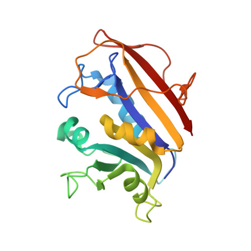
    Structural data are reported for the first examples of the tetrahydroquinazoline antifolate (6R,6S)-2,4-diamino-6-(1-indolinomethyl)-5,6,7,8-tetrahydroquinazoline (1) and its trimethoxy analogue (6R,6S)-2,4-diamino-6-(3',4',5'-trimethoxybenzyl)-5,6,7,8-tetrahydroquinazoline (2) as inhibitor complexes with dihydrofolate reductase (DHFR) from human (hDHFR) and Pneumocystis carinii (pcDHFR) sources. The indoline analogue (1) was crystallized as ternary complexes with NADPH and hDHFR (1.9 A resolution) and pcDHFR (2.3 A resolution), while the trimethoxy quinazoline analogue (2) was crystallized as a binary complex with hDHFR in two polymorphic rhombohedral R3 lattices: R3(1) to 1.8 A resolution and R3(2) to 2.0 A resolution. Structural analysis of these potent and selective DHFR-inhibitor complexes revealed preferential binding of the 6S-equatorial isomer in each structure. This configuration is similar to that of the natural tetrahydrofolate substrate; that is, 6S. These data also show that in both the hDHFR and pcDHFR ternary complexes with (1) the indoline ring is partially disordered, with two static conformations that differ between structures. These conformers also differ from that observed for the trimethoxybenzyl ring of tetrahydroquinazoline (2). There is also a correlation between the disorder of the flexible loop 23 and the disorder of the cofactor nicotinamide ribose ring in the pcDHFR-NADPH-(1) ternary complex. Comparison of the Toxoplasma gondii DHFR (tgDHFR) sequence with those of other DHFRs provides insight into the role of sequence and conformation in inhibitor-binding preferences which may aid in the design of novel antifolates with specific DHFR selectivity.


  • Organizational Affiliation

    Structural Biology Department, Hauptman-Woodward Medical Research Institute, 73 High Street, Buffalo, NY 14203, USA. cody@hwi.buffalo.edu


Macromolecules
Find similar proteins by:  (by identity cutoff)  |  3D Structure
Entity ID: 1
MoleculeChains Sequence LengthOrganismDetailsImage
Dihydrofolate reductase186Homo sapiensMutation(s): 0 
Gene Names: DHFR
EC: 1.5.1.3
UniProt & NIH Common Fund Data Resources
Find proteins for P00374 (Homo sapiens)
Explore P00374 
Go to UniProtKB:  P00374
PHAROS:  P00374
GTEx:  ENSG00000228716 
Entity Groups  
Sequence Clusters30% Identity50% Identity70% Identity90% Identity95% Identity100% Identity
UniProt GroupP00374
Sequence Annotations
Expand
  • Reference Sequence
Small Molecules
Ligands 2 Unique
IDChains Name / Formula / InChI Key2D Diagram3D Interactions
TQD
Query on TQD

Download Ideal Coordinates CCD File 
D [auth A](2R,6S)-6-{[methyl(3,4,5-trimethoxyphenyl)amino]methyl}-1,2,5,6,7,8-hexahydroquinazoline-2,4-diamine
C19 H29 N5 O3
JJWPLCQODKLEHY-JEOXALJRSA-N
SO4
Query on SO4

Download Ideal Coordinates CCD File 
B [auth A],
C [auth A]
SULFATE ION
O4 S
QAOWNCQODCNURD-UHFFFAOYSA-L
Experimental Data & Validation

Experimental Data

  • Method: X-RAY DIFFRACTION
  • Resolution: 2.50 Å
  • R-Value Free: 0.182 
  • R-Value Work: 0.165 
  • R-Value Observed: 0.165 
  • Space Group: H 3
Unit Cell:
Length ( Å )Angle ( ˚ )
a = 85.173α = 90
b = 85.173β = 90
c = 77.856γ = 120
Software Package:
Software NamePurpose
SCALEPACKdata scaling
PROTEINmodel building
PROFFTrefinement
PROTEINphasing

Structure Validation

View Full Validation Report


Ligand Structure Quality Assessment 


Entry History 

Deposition Data

Revision History  (Full details and data files)

  • Version 1.0: 2004-03-30
    Type: Initial release
  • Version 1.1: 2008-04-29
    Changes: Version format compliance
  • Version 1.2: 2011-07-13
    Changes: Version format compliance
  • Version 1.3: 2017-10-11
    Changes: Refinement description
  • Version 1.4: 2024-02-14
    Changes: Data collection, Database references, Derived calculations