1S2D

Purine 2'-Deoxyribosyl complex with arabinoside: Ribosylated Intermediate (AraA)


Experimental Data Snapshot

  • Method: X-RAY DIFFRACTION
  • Resolution: 2.10 Å
  • R-Value Free: 0.253 
  • R-Value Work: 0.229 
  • R-Value Observed: 0.229 

wwPDB Validation   3D Report Full Report


Ligand Structure Quality Assessment 


This is version 2.1 of the entry. See complete history


Literature

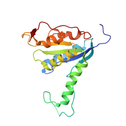
Structures of purine 2'-deoxyribosyltransferase, substrate complexes, and the ribosylated enzyme intermediate at 2.0 A resolution.

Anand, R.Kaminski, P.A.Ealick, S.E.

(2004) Biochemistry 43: 2384-2393

  • DOI: https://doi.org/10.1021/bi035723k
  • Primary Citation of Related Structures:  
    1S2D, 1S2G, 1S2I, 1S2L, 1S3F

  • PubMed Abstract: 

    The structure of class I N-deoxyribosyltransferase from Lactobacillus helveticus was determined by X-ray crystallography. Unlike class II N-deoxyribosyltransferases, which accept either purine or pyrimidine deoxynucleosides, class I enzymes are specific for purines as both the donor and acceptor base. Both class I and class II enzymes are highly specific for deoxynucleosides. The class I structure reveals similarities with the previously determined class II enzyme from Lactobacillus leichmanni [Armstrong, S. A., Cook, W. J., Short, S. A., and Ealick, S. E. (1996) Structure 4, 97-107]. The specificity of the class I enzyme for purine deoxynucleosides can be traced to a loop (residues 48-62), which shields the active site in the class II enzyme. In the class I enzyme, the purine base itself shields the active site from the solvent, while the smaller pyrimidine base cannot. The structure of the enzyme with a bound ribonucleoside shows that the nucleophilic oxygen atom of Glu101 hydrogen bonds to the O2' atom, rendering it unreactive and thus explaining the specificity for 2'-deoxynucleosides. The structure of a ribosylated enzyme intermediate reveals movements that occur during cleavage of the N-glycosidic bond. The structures of complexes with substrates and substrate analogues show that the purine base can bind in several different orientations, thus explaining the ability of the enzyme to catalyze alternate deoxyribosylation at the N3 or N7 position.


  • Organizational Affiliation

    Department of Chemistry and Chemical Biology, Cornell University, Ithaca, New York 14853, USA.


Macromolecules
Find similar proteins by:  (by identity cutoff)  |  3D Structure
Entity ID: 1
MoleculeChains Sequence LengthOrganismDetailsImage
Nucleoside 2-deoxyribosyltransferase
A, B, C
167Lactobacillus helveticusMutation(s): 0 
Gene Names: ptdndtAntd_1BC335_1936BCM45_08885BDKNPLJD_00991DM475_00825GDZ32_10450LH5_01102
EC: 2.4.2.6
UniProt
Find proteins for Q8RLY5 (Lactobacillus helveticus)
Explore Q8RLY5 
Go to UniProtKB:  Q8RLY5
Entity Groups  
Sequence Clusters30% Identity50% Identity70% Identity90% Identity95% Identity100% Identity
UniProt GroupQ8RLY5
Sequence Annotations
Expand
  • Reference Sequence
Experimental Data & Validation

Experimental Data

  • Method: X-RAY DIFFRACTION
  • Resolution: 2.10 Å
  • R-Value Free: 0.253 
  • R-Value Work: 0.229 
  • R-Value Observed: 0.229 
  • Space Group: P 43 21 2
Unit Cell:
Length ( Å )Angle ( ˚ )
a = 79.587α = 90
b = 79.587β = 90
c = 184.973γ = 90
Software Package:
Software NamePurpose
CNSrefinement
DENZOdata reduction
SCALEPACKdata scaling
CNSphasing

Structure Validation

View Full Validation Report


Ligand Structure Quality Assessment 


Entry History 

Deposition Data

Revision History  (Full details and data files)

  • Version 1.0: 2004-03-30
    Type: Initial release
  • Version 1.1: 2008-04-29
    Changes: Version format compliance
  • Version 1.2: 2011-07-13
    Changes: Derived calculations, Version format compliance
  • Version 2.0: 2020-07-01
    Changes: Advisory, Atomic model, Data collection, Database references, Derived calculations, Non-polymer description, Polymer sequence, Source and taxonomy, Structure summary
  • Version 2.1: 2020-07-29
    Type: Remediation
    Reason: Carbohydrate remediation
    Changes: Derived calculations