Structural insight into arginine degradation by arginine deiminase, an antibacterial and parasite drug target.
Galkin, A., Kulakova, L., Sarikaya, E., Lim, K., Howard, A., Herzberg, O.(2004) J Biol Chem 279: 14001-14008
- PubMed: 14701825
- DOI: https://doi.org/10.1074/jbc.M313410200
- Primary Citation of Related Structures:
1RXX - PubMed Abstract:
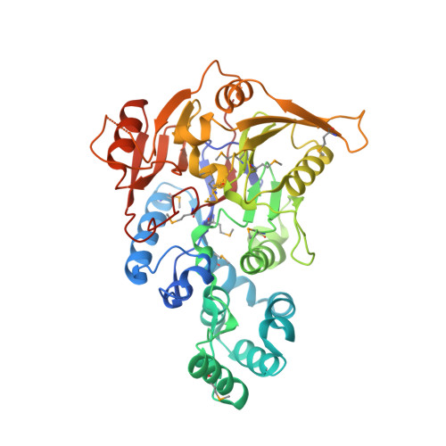
l-Arginine deiminase (ADI) catalyzes the irreversible hydrolysis of arginine to citrulline and ammonia. ADI is involved in the first step of the most widespread anaerobic route of arginine degradation. ADI, missing in high eukaryotes, is a potential antimicrobial and antiparasitic drug target. We have determined the crystal structure of ADI from Pseudomonas aeruginosa by the multi-wavelength anomalous diffraction method at 2.45 A resolution. The structure exhibits similarity to other arginine-modifying or substituted arginine-modifying enzymes such as dimethylarginine dimethylaminohydrolase (DDAH), arginine:glycine amidinotransferase, and arginine:inosamine-phosphate amidinotransferase, despite the lack of significant amino acid sequence homology to these enzymes. The similarity spans a core domain comprising five betabetaalphabeta motifs arranged in a circle around a 5-fold pseudosymmetry axis. ADI contains an additional alpha-helical domain of novel topology inserted between the first and the second betabetaalphabeta modules. A catalytic triad, Cys-His-Glu/Asp (arranged in a different manner from that of the thiol proteases), seen in the other arginine-modifying enzymes is also conserved in ADI, as well as many other residues involved in substrate binding. Based on this conservation pattern and the assumption that the substrate binding mode is similar to that of DDAH, an ADI catalytic mechanism is proposed. The main players are Cys-406, which mounts the nucleophilic attack on the carbon atom of the guanidinium group of arginine, and His-278, which serves as a general base.
Organizational Affiliation:
Center for Advanced Research In Biotechnology, University of Maryland Biotechnology Institute, Rockville, Maryland 20850, USA.