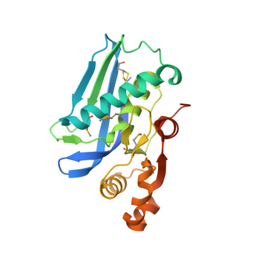
The catalytic domain of Escherichia coli Lon protease has a unique fold and a Ser-Lys dyad in the active site
Botos, I., Melnikov, E.E., Cherry, S., Tropea, J.E., Khalatova, A.G., Rasulova, F., Dauter, Z., Maurizi, M.R., Rotanova, T.V., Wlodawer, A., Gustchina, A.(2004) J Biol Chem 279: 8140-8148
- PubMed: 14665623
- DOI: https://doi.org/10.1074/jbc.M312243200
- Primary Citation of Related Structures:
1RR9, 1RRE - PubMed Abstract:
ATP-dependent Lon protease degrades specific short-lived regulatory proteins as well as defective and abnormal proteins in the cell. The crystal structure of the proteolytic domain (P domain) of the Escherichia coli Lon has been solved by single-wavelength anomalous dispersion and refined at 1.75-A resolution. The P domain was obtained by chymotrypsin digestion of the full-length, proteolytically inactive Lon mutant (S679A) or by expression of a recombinant construct encoding only this domain. The P domain has a unique fold and assembles into hexameric rings that likely mimic the oligomerization state of the holoenzyme. The hexamer is dome-shaped, with the six N termini oriented toward the narrower ring surface, which is thus identified as the interface with the ATPase domain in full-length Lon. The catalytic sites lie in a shallow concavity on the wider distal surface of the hexameric ring and are connected to the proximal surface by a narrow axial channel with a diameter of approximately 18 A. Within the active site, the proximity of Lys(722) to the side chain of the mutated Ala(679) and the absence of other potential catalytic side chains establish that Lon employs a Ser(679)-Lys(722) dyad for catalysis. Alignment of the P domain catalytic pocket with those of several Ser-Lys dyad peptide hydrolases provides a model of substrate binding, suggesting that polypeptides are oriented in the Lon active site to allow nucleophilic attack by the serine hydroxyl on the si-face of the peptide bond.
Organizational Affiliation:
Macromolecular Crystallography Laboratory, National Cancer Institute at Frederick, Frederick, Maryland 21702-1201, USA.