1RCB

CRYSTAL STRUCTURE OF HUMAN RECOMBINANT INTERLEUKIN-4 AT 2.25 ANGSTROMS RESOLUTION


Experimental Data Snapshot

  • Method: X-RAY DIFFRACTION
  • Resolution: 2.25 Å
  • R-Value Observed: 0.218 

wwPDB Validation   3D Report Full Report


This is version 1.2 of the entry. See complete history


Literature

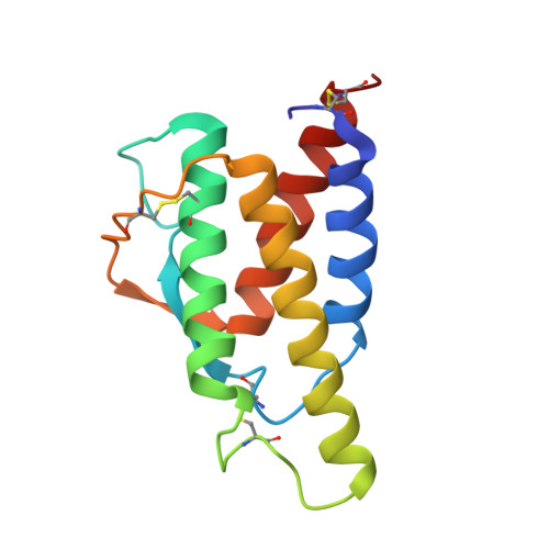
Crystal structure of human recombinant interleukin-4 at 2.25 A resolution.

Wlodawer, A.Pavlovsky, A.Gustchina, A.

(1992) FEBS Lett 309: 59-64

  • DOI: https://doi.org/10.1016/0014-5793(92)80739-4
  • Primary Citation of Related Structures:  
    1RCB

  • PubMed Abstract: 

    The crystal structure of human recombinant interleukin-4 (IL-4) has been solved by multiple isomorphous replacement, and refined to an R factor of 0.218 at 2.25 A resolution. The molecule is a left-handed four-helix bundle with a short stretch of beta sheet. The structure bears close resemblance to other cytokines such as granulocyte-macrophage colony stimulating factor (GM-CSF). Although no sequence similarity of IL-4 to GM-CSF and other related cytokines has been previously postulated, structure-based alignment of IL-4 and GM-CSF revealed that the core of the molecules, including large parts of all four helices and extending over half of the molecule, has 30% sequence identity. This may have identified regions which are not only important to maintain structure, but could also play a role in receptor binding.


  • Organizational Affiliation

    Macromolucar Structure Laboratory, NCI-Fredrick Cancer Research and Development Center, MD 21702.


Macromolecules
Find similar proteins by:  (by identity cutoff)  |  3D Structure
Entity ID: 1
MoleculeChains Sequence LengthOrganismDetailsImage
INTERLEUKIN-4129Homo sapiensMutation(s): 0 
UniProt & NIH Common Fund Data Resources
Find proteins for P05112 (Homo sapiens)
Explore P05112 
Go to UniProtKB:  P05112
PHAROS:  P05112
GTEx:  ENSG00000113520 
Entity Groups  
Sequence Clusters30% Identity50% Identity70% Identity90% Identity95% Identity100% Identity
UniProt GroupP05112
Sequence Annotations
Expand
  • Reference Sequence
Experimental Data & Validation

Experimental Data

  • Method: X-RAY DIFFRACTION
  • Resolution: 2.25 Å
  • R-Value Observed: 0.218 
  • Space Group: P 41 21 2
Unit Cell:
Length ( Å )Angle ( ˚ )
a = 91.8α = 90
b = 91.8β = 90
c = 46.4γ = 90
Software Package:
Software NamePurpose
PROLSQrefinement

Structure Validation

View Full Validation Report


Entry History 

Deposition Data

Revision History  (Full details and data files)

  • Version 1.0: 1993-10-31
    Type: Initial release
  • Version 1.1: 2008-03-21
    Changes: Version format compliance
  • Version 1.2: 2011-07-13
    Changes: Version format compliance