1RB8

The phiX174 DNA binding protein J in two different capsid environments.


Experimental Data Snapshot

  • Method: X-RAY DIFFRACTION
  • Resolution: 3.50 Å
  • R-Value Free: 0.236 
  • R-Value Work: 0.234 
  • R-Value Observed: 0.234 

wwPDB Validation   3D Report Full Report


Ligand Structure Quality Assessment 


This is version 2.1 of the entry. See complete history


Literature

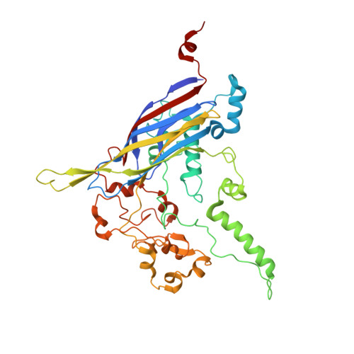
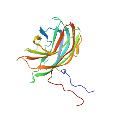
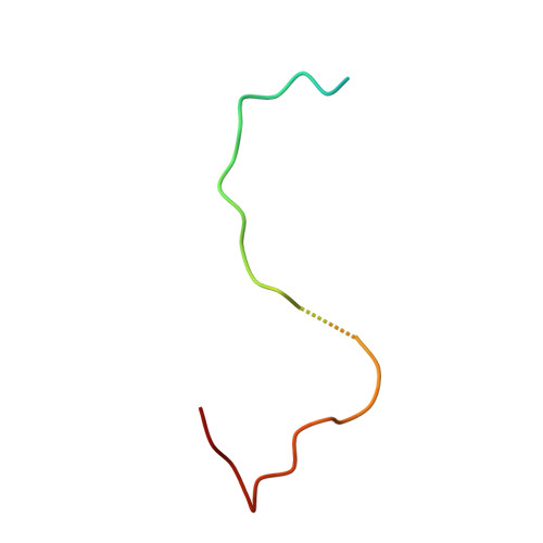
The phiX174 Protein J Mediates DNA Packaging and Viral Attachment to Host Cells.

Bernal, R.A.Hafenstein, S.Esmeralda, R.Fane, B.A.Rossmann, M.G.

(2004) J Mol Biol 337: 1109-1122

  • DOI: https://doi.org/10.1016/j.jmb.2004.02.033
  • Primary Citation of Related Structures:  
    1RB8

  • PubMed Abstract: 

    Packaging of viral genomes into their respective capsids requires partial neutralization of the highly negatively charged RNA or DNA. Many viruses, including the Microviridae bacteriophages phiX174, G4, and alpha3, have solved this problem by coding for a highly positively charged nucleic acid-binding protein that is packaged along with the genome. The phiX174 DNA-binding protein, J, is 13 amino acid residues longer than the alpha3 and G4 J proteins by virtue of an additional nucleic acid-binding domain at the amino terminus. Chimeric phiX174 particles containing the smaller DNA-binding protein cannot be generated due to procapsid instability during DNA packaging. However, chimeric alpha3 and G4 phages, containing the phiX174 DNA-binding protein in place of the endogenous J protein, assemble and are infectious, but are less dense than the respective wild-type species. In addition, host cell attachment and native gel migration assays indicate surface variations of these viruses that are controlled by the nature of the J protein. The structure of alpha3 packaged with phiX174 J protein was determined to 3.5A resolution and compared with the previously determined structures of phiX174 and alpha3. The structures of the capsid and spike proteins in the chimeric particle remain unchanged within experimental error when compared to the wild-type alpha3 virion proteins. The amino-terminal region of the phiX174 J protein, which is missing from wild-type alpha3 virions, is mostly disordered in the alpha3 chimera. The differences observed between solution properties of wild-type phiX174, wild-type alpha3, and alpha3 chimera, including their ability to attach to host cells, correlates with the degree of order in the amino-terminal domain of the J protein. When ordered, this domain binds to the interior of the viral capsid and, thus, might control the flexibility of the capsid. In addition, the properties of the phiX174 J protein in the chimera and the results of mutational analyses suggest that an evolutionary correlation may exist between the size of the J protein and the stoichiometry of the DNA pilot protein H, required in the initial stages of infection. Hence, the function of the J protein is to facilitate DNA packaging, as well as to mediate surface properties such as cell attachment and infection.


  • Organizational Affiliation

    Department of Biological Sciences, Purdue University, Lilly Hall, 915 W State Street, West Lafayette, IN 47907-2054, USA.


Macromolecules

Find similar proteins by:  (by identity cutoff)  |  3D Structure
Entity ID: 1
MoleculeChains Sequence LengthOrganismDetailsImage
Capsid proteinA [auth F]431Escherichia phage alpha3Mutation(s): 0 
UniProt
Find proteins for P08767 (Escherichia phage alpha3)
Explore P08767 
Go to UniProtKB:  P08767
Entity Groups  
Sequence Clusters30% Identity50% Identity70% Identity90% Identity95% Identity100% Identity
UniProt GroupP08767
Sequence Annotations
Expand
  • Reference Sequence
Find similar proteins by:  (by identity cutoff)  |  3D Structure
Entity ID: 2
MoleculeChains Sequence LengthOrganismDetailsImage
Major spike proteinB [auth G]187Escherichia phage alpha3Mutation(s): 0 
UniProt
Find proteins for P31281 (Escherichia phage alpha3)
Explore P31281 
Go to UniProtKB:  P31281
Entity Groups  
Sequence Clusters30% Identity50% Identity70% Identity90% Identity95% Identity100% Identity
UniProt GroupP31281
Sequence Annotations
Expand
  • Reference Sequence
Find similar proteins by:  (by identity cutoff)  |  3D Structure
Entity ID: 3
MoleculeChains Sequence LengthOrganismDetailsImage
Small core proteinC [auth J]37Sinsheimervirus phiX174Mutation(s): 0 
Gene Names: J
UniProt
Find proteins for P69592 (Enterobacteria phage phiX174)
Explore P69592 
Go to UniProtKB:  P69592
Entity Groups  
Sequence Clusters30% Identity50% Identity70% Identity90% Identity95% Identity100% Identity
UniProt GroupP69592
Sequence Annotations
Expand
  • Reference Sequence

Find similar nucleic acids by:  Sequence   |   3D Structure  

Entity ID: 4
MoleculeChains LengthOrganismImage
DNA (5'-D(P*CP*AP*AP*A)-3')D [auth X]4N/A
Sequence Annotations
Expand
  • Reference Sequence
Experimental Data & Validation

Experimental Data

  • Method: X-RAY DIFFRACTION
  • Resolution: 3.50 Å
  • R-Value Free: 0.236 
  • R-Value Work: 0.234 
  • R-Value Observed: 0.234 
  • Space Group: H 3
Unit Cell:
Length ( Å )Angle ( ˚ )
a = 295.143α = 90
b = 295.143β = 90
c = 678.235γ = 120
Software Package:
Software NamePurpose
CNSrefinement
DENZOdata reduction
SnBphasing
ENVELOPEphasing

Structure Validation

View Full Validation Report


Ligand Structure Quality Assessment 


Entry History 

Deposition Data

Revision History  (Full details and data files)

  • Version 1.0: 2004-04-13
    Type: Initial release
  • Version 1.1: 2008-04-01
    Changes: Version format compliance
  • Version 1.2: 2011-07-13
    Changes: Version format compliance
  • Version 1.3: 2017-10-11
    Changes: Refinement description
  • Version 1.4: 2018-01-31
    Changes: Experimental preparation
  • Version 2.0: 2023-04-19
    Type: Remediation
    Changes: Advisory, Atomic model, Data collection, Database references, Derived calculations, Other, Refinement description
  • Version 2.1: 2024-04-03
    Changes: Data collection, Refinement description