1QKQ

CHARCOT-LEYDEN CRYSTAL PROTEIN - MANNOSE COMPLEX


Experimental Data Snapshot

  • Method: X-RAY DIFFRACTION
  • Resolution: 1.80 Å
  • R-Value Free: 0.251 
  • R-Value Work: 0.236 
  • R-Value Observed: 0.236 

wwPDB Validation   3D Report Full Report


This is version 1.4 of the entry. See complete history


Literature

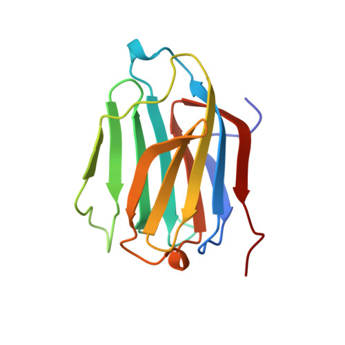
Selective recognition of mannose by the human eosinophil Charcot-Leyden crystal protein (galectin-10): a crystallographic study at 1.8 A resolution.

Swaminathan, G.J.Leonidas, D.D.Savage, M.P.Ackerman, S.J.Acharya, K.R.

(1999) Biochemistry 38: 13837-13843

  • DOI: https://doi.org/10.1021/bi990756e
  • Primary Citation of Related Structures:  
    1QKQ

  • PubMed Abstract: 

    The role(s) of the eosinophil Charcot-Leyden crystal (CLC) protein in eosinophil or basophil function or associated inflammatory processes is yet to be established. Although the CLC protein has been reported to exhibit weak lysophospholipase activity, it shows virtually no sequence homology to any known member of this family of enzymes. The X-ray crystal structure of the CLC protein is very similar to the structure of the galectins, members of a beta-galactoside-specific animal lectin family, including a partially conserved galectin carbohydrate recognition domain (CRD). In the absence of any known natural carbohydrate ligand for this protein, the functional role of the CLC protein (galectin-10) has remained speculative. Here we describe structural studies on the carbohydrate binding properties of the CLC protein and report the first structure of a carbohydrate in complex with the protein. Interestingly, the CLC protein demonstrates no affinity for beta-galactosides and binds mannose in a manner very different from those of other related galectins that have been shown to bind lactosamine. The partial conservation of residues involved in carbohydrate binding led to significant changes in the topology and chemical nature of the CRD, and has implications for carbohydrate recognition by the CLC protein in vivo and its functional role in the biology of inflammation.


  • Organizational Affiliation

    Department of Biology and Biochemistry, University of Bath, Claverton Down, U.K.


Macromolecules
Find similar proteins by:  (by identity cutoff)  |  3D Structure
Entity ID: 1
MoleculeChains Sequence LengthOrganismDetailsImage
EOSINOPHIL LYSOPHOSPHOLIPASE142Homo sapiensMutation(s): 0 
EC: 3.1.1.5
UniProt & NIH Common Fund Data Resources
Find proteins for Q05315 (Homo sapiens)
Explore Q05315 
Go to UniProtKB:  Q05315
PHAROS:  Q05315
GTEx:  ENSG00000105205 
Entity Groups  
Sequence Clusters30% Identity50% Identity70% Identity90% Identity95% Identity100% Identity
UniProt GroupQ05315
Sequence Annotations
Expand
  • Reference Sequence
Small Molecules
Ligands 1 Unique
IDChains Name / Formula / InChI Key2D Diagram3D Interactions
MAN
Query on MAN

Download Ideal Coordinates CCD File 
B [auth A]alpha-D-mannopyranose
C6 H12 O6
WQZGKKKJIJFFOK-PQMKYFCFSA-N
Experimental Data & Validation

Experimental Data

  • Method: X-RAY DIFFRACTION
  • Resolution: 1.80 Å
  • R-Value Free: 0.251 
  • R-Value Work: 0.236 
  • R-Value Observed: 0.236 
  • Space Group: P 65 2 2
Unit Cell:
Length ( Å )Angle ( ˚ )
a = 49.6α = 90
b = 49.6β = 90
c = 263.13γ = 120
Software Package:
Software NamePurpose
CNSrefinement
DENZOdata reduction
SCALEPACKdata scaling
AMoREphasing

Structure Validation

View Full Validation Report


Entry History 

Deposition Data

Revision History  (Full details and data files)

  • Version 1.0: 2000-01-18
    Type: Initial release
  • Version 1.1: 2012-08-29
    Changes: Atomic model, Database references, Derived calculations, Non-polymer description, Other, Refinement description, Structure summary, Version format compliance
  • Version 1.2: 2019-09-18
    Changes: Data collection, Database references
  • Version 1.3: 2020-07-29
    Type: Remediation
    Reason: Carbohydrate remediation
    Changes: Data collection, Derived calculations, Structure summary
  • Version 1.4: 2023-12-13
    Changes: Data collection, Database references, Refinement description, Structure summary