Effect of N-terminal and Met23 Mutations on the Structure and Dynamics of Onconase
Gorbatyuk, V.Y., Tsai, C.K., Chang, C.F., Huang, T.H.(2004) J Biol Chem 279: 5772-5780
- PubMed: 14645226
- DOI: https://doi.org/10.1074/jbc.M311233200
- Primary Citation of Related Structures:
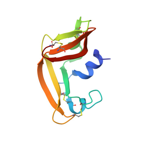
1PU3 - PubMed Abstract:
Onconase (rONC), otherwise known as ranpirnase or P-30 protein, which was initially purified from extracts of Rana pipiens oocytes and early embryos, exhibits anticancer activity both in vitro and in vivo and is in phase III clinical trials for tumor therapy. We have determined the solution NMR structure of a recombinant onconase with Met(-1), Gln1, and Leu23 residues (M-1, Q1, M23L)rONC. The 20 best solution structures had a backbone root mean square deviation of 0.41 +/- 0.09 A with respect to the average structure. The energy-minimized average NMR structure had a backbone root mean square deviation of 0.72 A from the x-ray crystallographic structure of native onconase; however, the orientation of the N-terminal residue in the two structures was very different. Comparison of the 15N HSQC spectrum of (M-1, Q1, M23L)rONC with that of a mutant E1S-rONC, which is identical to the nONC except with the N-terminal pyroglutamyl residue replaced by Ser, showed that N-terminal and residue 23 mutations induced structural changes in regions beyond the mutation sites. Model-free analysis of the backbone amide 15N-T1, 15N-T2, and 15N-1H NOE relaxation data for (M-1, Q1, M23L)rONC and E1S-rONC revealed that the E1S-rONC molecule showed very little flexibility, whereas (M-1, Q1, M23L)rONC exhibited substantial flexibility, which may account for the previously observed reduced stability and increased protease susceptibility. The alpha1 helix and beta-sheets of (M-1, Q1, M23L)rONC displayed bending motions. These data provided strong evidence for the presence of an N-terminal hydrogen bond network in E1S-rONC, but not in (M-1, Q1, M23L)rONC.
Organizational Affiliation:
Institute of Biomedical Sciences, Academia Sinica, Nankang, Taipei 11529, Taiwan, Republic of China.