


1PMH
Crystal structure of Caldicellulosiruptor saccharolyticus CBM27-1 in complex with mannohexaose
- PDB DOI: https://doi.org/10.2210/pdb1PMH/pdb
- Classification: HYDROLASE
- Organism(s): Caldicellulosiruptor saccharolyticus
- Expression System: Escherichia coli
- Mutation(s): Yes
- Deposited: 2003-06-11 Released: 2004-06-22
Experimental Data Snapshot
- Method: X-RAY DIFFRACTION
- Resolution: 1.06 Å
- R-Value Free: 0.173
- R-Value Work: 0.141
- R-Value Observed: 0.143
wwPDB Validation 3D Report Full Report

This is version 2.0 of the entry. See complete history.

Macromolecules
Find similar proteins by:
(by identity cutoff) | 3D Structure
Entity ID: 1 | |||||
|---|---|---|---|---|---|
| Molecule | Chains | Sequence Length | Organism | Details | Image |
| beta-1,4-mannanase | A [auth X] | 185 | Caldicellulosiruptor saccharolyticus | Mutation(s): 2 Gene Names: manA EC: 3.2.1.78 | 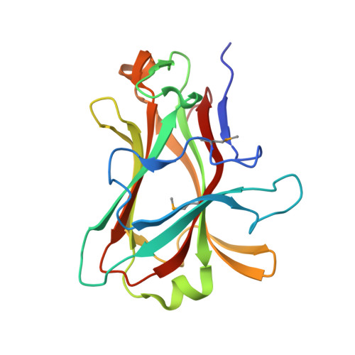 |
UniProt | |||||
Find proteins for P77847 (Caldicellulosiruptor saccharolyticus) Explore P77847 Go to UniProtKB: P77847 | |||||
Entity Groups | |||||
| Sequence Clusters | 30% Identity50% Identity70% Identity90% Identity95% Identity100% Identity | ||||
| UniProt Group | P77847 | ||||
Sequence AnnotationsExpand | |||||
| |||||
Oligosaccharides
Entity ID: 2 | |||||
|---|---|---|---|---|---|
| Molecule | Chains | Length | 2D Diagram | Glycosylation | 3D Interactions |
| beta-D-mannopyranose-(1-4)-beta-D-mannopyranose-(1-4)-beta-D-mannopyranose-(1-4)-beta-D-mannopyranose-(1-4)-beta-D-mannopyranose-(1-4)-alpha-D-mannopyranose | B [auth A] | 6 |  | N/A | |
Glycosylation Resources | |||||
GlyTouCan: G39448BE GlyCosmos: G39448BE GlyGen: G39448BE | |||||
Small Molecules
| Ligands 2 Unique | |||||
|---|---|---|---|---|---|
| ID | Chains | Name / Formula / InChI Key | 2D Diagram | 3D Interactions | |
| EDO Query on EDO | D [auth X], E [auth X], F [auth X], G [auth X] | 1,2-ETHANEDIOL C2 H6 O2 LYCAIKOWRPUZTN-UHFFFAOYSA-N |  | ||
| CA Query on CA | C [auth X] | CALCIUM ION Ca BHPQYMZQTOCNFJ-UHFFFAOYSA-N |  | ||
| Modified Residues 1 Unique | |||||
|---|---|---|---|---|---|
| ID | Chains | Type | Formula | 2D Diagram | Parent |
| MSE Query on MSE | A [auth X] | L-PEPTIDE LINKING | C5 H11 N O2 Se |  | MET |
Experimental Data & Validation
Experimental Data
- Method: X-RAY DIFFRACTION
- Resolution: 1.06 Å
- R-Value Free: 0.173
- R-Value Work: 0.141
- R-Value Observed: 0.143
- Space Group: P 21 21 21
Unit Cell:
| Length ( Å ) | Angle ( ˚ ) |
|---|---|
| a = 38.281 | α = 90 |
| b = 45.7 | β = 90 |
| c = 110.113 | γ = 90 |
| Software Name | Purpose |
|---|---|
| REFMAC | refinement |
| MAR345 | data collection |
| XDS | data scaling |
| SOLVE | phasing |
| RESOLVE | phasing |
Entry History
Deposition Data
- Released Date: 2004-06-22 Deposition Author(s): Roske, Y., Sunna, A., Heinemann, U.
Revision History (Full details and data files)
- Version 1.0: 2004-06-22
Type: Initial release - Version 1.1: 2008-04-29
Changes: Version format compliance - Version 1.2: 2011-07-13
Changes: Version format compliance - Version 1.3: 2017-10-11
Changes: Refinement description - Version 2.0: 2020-07-29
Type: Remediation
Reason: Carbohydrate remediation
Changes: Atomic model, Data collection, Database references, Derived calculations, Structure summary
