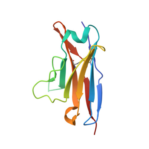
Structural basis of light chain amyloidogenicity: comparison of the thermodynamic properties, fibrillogenic potential and tertiary structural features of four V(lambda)6 proteins
Wall, J.S., Gupta, V., Wilkerson, M., Schell, M., Loris, R., Adams, P., Solomon, A., Stevens, F., Dealwis, C.(2004) J Mol Recognit 17: 323-331
- PubMed: 15227639
- DOI: https://doi.org/10.1002/jmr.681
- Primary Citation of Related Structures:
1PEW, 1PW3 - PubMed Abstract:
Primary (AL) amyloidosis results from the pathologic deposition of monoclonal light chains as amyloid fibrils. Studies of recombinant-derived variable region (VL) fragments of these proteins have shown an inverse relationship between thermodynamic stability and fibrillogenic potential. Further, ionic interactions within the VL domain were predicted to influence the kinetics of light chain fibrillogenicity, as evidenced from our analyses of a relatively stable Vlambda6 protein (Jto) with a long range electrostatic interaction between Asp and Arg side chains at position 29 and 68, respectively, and an unstable, highly fibrillogenic Vlambda6 protein (Wil) that had neutral amino acids at these locations. To test this hypothesis, we have generated two Jto-related mutants designed to disrupt the interaction between Asp 29 and Arg 68 (JtoD29A and JtoR68S). Although the thermodynamic stabilities of unfolding for these two molecules were identical, they exhibited very different kinetics of fibril formation: the rate of JtoD29A fibrillogenesis was slow and comparable to the parent molecule, whereas that of JtoR68S was significantly faster. High-resolution X-ray diffraction analyses of crystals prepared from the two mutants having the same space group and unit cell dimensions revealed no significant main-chain conformational changes. However, several notable side-chain alterations were observed in JtoR68S, as compared with JtoD29A, that resulted in the solvent exposure of a greater hydrophobic surface and modifications in the electrostatic potential surface. We posit that these differences contributed to the enhanced fibrillogenic potential of the Arg 68 mutant, since both Jto mutants lacked the intrachain ionic interaction and were equivalently unstable. The information gleaned from our studies has provided insight into structural parameters that in addition to overall thermodynamic stability, contribute to the fibril forming propensity of immunoglobulin light chains.
Organizational Affiliation:
Human Immunology and Cancer Program, Department of Medicine, University of Tennessee Graduate School of Medicine, Knoxville, Tennessee, USA.