1PB3

Sites of binding and orientation in a four location model for protein stereospecificity.


Experimental Data Snapshot

  • Method: X-RAY DIFFRACTION
  • Resolution: 1.70 Å
  • R-Value Free: 0.216 
  • R-Value Work: 0.194 
  • R-Value Observed: 0.194 

wwPDB Validation   3D Report Full Report


This is version 1.4 of the entry. See complete history


Literature

Sites of Binding and Orientation in a Four-Location Model for Protein Stereospecificity.

Mesecar, A.D.Koshland Jr., D.E.

(2000) IUBMB Life 49: 457-466

  • DOI: https://doi.org/10.1080/152165400410326
  • Primary Citation of Related Structures:  
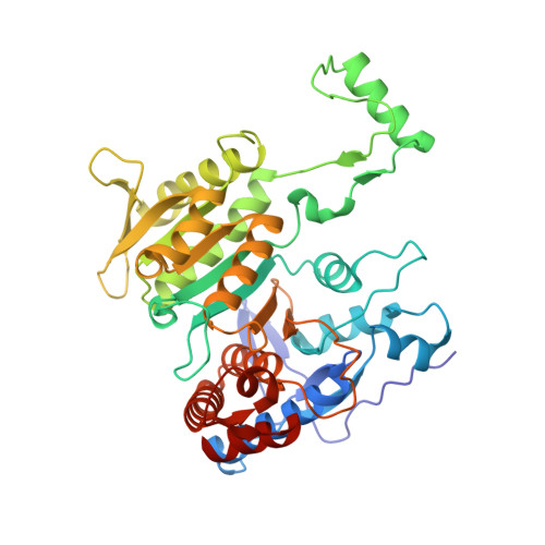
    1PB3

  • PubMed Abstract: 

    The stereospecificity of the enzyme isocitrate dehydrogenase was examined by steady-state kinetics and x-ray crystallography. The enzyme has the intriguing property that the apoenzyme in the absence of divalent metal showed a selectivity for the inactive l-enantiomer of the substrate isocitrate, whereas the enzyme containing magnesium showed selectivity for the physiologically active d-enantiomer. The hydrogen atom on the C2 carbon that is transferred during the reaction was, in both the d- and l-isocitrate complexes, in an orientation very close to that expected for delivery of a hydride ion to the cosubstrate NADP+. The beta-carboxylate that is eliminated as a CO2 molecule during the reaction occupied the same site on the protein in both the d- and l-isocitrate complexes. In addition, the C3 carbon was in the same protein site in both the d- and l-enantiomers. Only the fourth group, the OH atom, was in a very different position in the apo enzyme and in the metal-containing complexes. A four-location model is necessary to explain the enantiomeric specificity of IDH in contrast to the conventional three-point attachment model. The thermodynamic and kinetic ramifications of this model are explored.


  • Organizational Affiliation

    Center for Pharmaceutical Biotechnology and the Department of Medicinal Chemistry and Pharmacognosy, University of Illinois at Chicago, 60607, USA. mesecar@uic.edu


Macromolecules
Find similar proteins by:  (by identity cutoff)  |  3D Structure
Entity ID: 1
MoleculeChains Sequence LengthOrganismDetailsImage
Isocitrate dehydrogenase [NADP]416Escherichia coliMutation(s): 0 
Gene Names: ICD OR ICDA OR ICDE OR B1136
EC: 1.1.1.42
UniProt
Find proteins for P08200 (Escherichia coli (strain K12))
Explore P08200 
Go to UniProtKB:  P08200
Entity Groups  
Sequence Clusters30% Identity50% Identity70% Identity90% Identity95% Identity100% Identity
UniProt GroupP08200
Sequence Annotations
Expand
  • Reference Sequence
Experimental Data & Validation

Experimental Data

  • Method: X-RAY DIFFRACTION
  • Resolution: 1.70 Å
  • R-Value Free: 0.216 
  • R-Value Work: 0.194 
  • R-Value Observed: 0.194 
  • Space Group: P 43 21 2
Unit Cell:
Length ( Å )Angle ( ˚ )
a = 103.669α = 90
b = 103.669β = 90
c = 150.124γ = 90
Software Package:
Software NamePurpose
CNSrefinement
DENZOdata reduction
SCALEPACKdata scaling
EPMRphasing

Structure Validation

View Full Validation Report


Entry History 

Deposition Data

Revision History  (Full details and data files)

  • Version 1.0: 2003-06-17
    Type: Initial release
  • Version 1.1: 2008-04-29
    Changes: Version format compliance
  • Version 1.2: 2011-07-13
    Changes: Derived calculations, Version format compliance
  • Version 1.3: 2018-01-31
    Changes: Experimental preparation
  • Version 1.4: 2024-02-14
    Changes: Data collection, Database references, Derived calculations