1P9P

The Crystal Structure of a M1G37 tRNA Methyltransferase, TrmD


Experimental Data Snapshot

  • Method: X-RAY DIFFRACTION
  • Resolution: 2.50 Å
  • R-Value Free: 0.247 
  • R-Value Work: 0.196 

wwPDB Validation   3D Report Full Report


Ligand Structure Quality Assessment 


This is version 1.5 of the entry. See complete history


Literature

Insights into Catalysis by a Knotted TrmD tRNA Methyltransferase.

Elkins, P.A.Watts, J.M.Zalacain, M.Van Thiel, A.Vitaszka, P.R.Redlak, M.Andraos-Selim, C.Rastinejad, F.Holmes, W.M.

(2003) J Mol Biol 333: 931-949

  • DOI: https://doi.org/10.1016/j.jmb.2003.09.011
  • Primary Citation of Related Structures:  
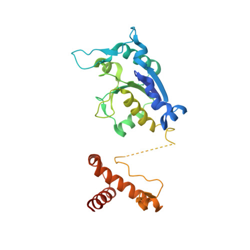
    1P9P

  • PubMed Abstract: 

    The crystal structure of Escherichia coli tRNA (guanosine-1) methyltransferase (TrmD) complexed with S-adenosyl homocysteine (AdoHcy) has been determined at 2.5A resolution. TrmD, which methylates G37 of tRNAs containing the sequence G36pG37, is a homo-dimer. Each monomer consists of a C-terminal domain connected by a flexible linker to an N-terminal AdoMet-binding domain. The two bound AdoHcy moieties are buried at the bottom of deep clefts. The dimer structure appears integral to the formation of the catalytic center of the enzyme and this arrangement strongly suggests that the anticodon loop of tRNA fits into one of these clefts for methyl transfer to occur. In addition, adjacent hydrophobic sites in the cleft delineate a defined pocket, which may accommodate the GpG sequence during catalysis. The dimer contains two deep trefoil peptide knots and a peptide loop extending from each knot embraces the AdoHcy adenine ring. Mutational analyses demonstrate that the knot is important for AdoMet binding and catalytic activity, and that the C-terminal domain is not only required for tRNA binding but plays a functional role in catalytic activity.


  • Organizational Affiliation

    GlaxoSmithKline, 709 Swedeland Road, UE0447, King of Prussia, PA 19406, USA.


Macromolecules
Find similar proteins by:  (by identity cutoff)  |  3D Structure
Entity ID: 1
MoleculeChains Sequence LengthOrganismDetailsImage
tRNA (Guanine-N(1)-)-methyltransferase261Escherichia coli K-12Mutation(s): 0 
Gene Names: TRMD OR B2607 OR C3128 OR Z3901 OR ECS3470 OR SF2667
EC: 2.1.1.31
UniProt
Find proteins for P0A873 (Escherichia coli (strain K12))
Explore P0A873 
Go to UniProtKB:  P0A873
Entity Groups  
Sequence Clusters30% Identity50% Identity70% Identity90% Identity95% Identity100% Identity
UniProt GroupP0A873
Sequence Annotations
Expand
  • Reference Sequence
Small Molecules
Ligands 1 Unique
IDChains Name / Formula / InChI Key2D Diagram3D Interactions
SAH
Query on SAH

Download Ideal Coordinates CCD File 
B [auth A]S-ADENOSYL-L-HOMOCYSTEINE
C14 H20 N6 O5 S
ZJUKTBDSGOFHSH-WFMPWKQPSA-N
Binding Affinity Annotations 
IDSourceBinding Affinity
SAH Binding MOAD:  1P9P Kd: 68 (nM) from 1 assay(s)
Experimental Data & Validation

Experimental Data

  • Method: X-RAY DIFFRACTION
  • Resolution: 2.50 Å
  • R-Value Free: 0.247 
  • R-Value Work: 0.196 
  • Space Group: P 32 2 1
Unit Cell:
Length ( Å )Angle ( ˚ )
a = 97.255α = 90
b = 97.255β = 90
c = 62.661γ = 120
Software Package:
Software NamePurpose
HKL-2000data collection
SCALEPACKdata scaling
SOLVEphasing
CNXrefinement
HKL-2000data reduction

Structure Validation

View Full Validation Report


Ligand Structure Quality Assessment 


Entry History 

Deposition Data

Revision History  (Full details and data files)

  • Version 1.0: 2004-05-18
    Type: Initial release
  • Version 1.1: 2008-04-29
    Changes: Version format compliance
  • Version 1.2: 2011-07-13
    Changes: Derived calculations, Version format compliance
  • Version 1.3: 2011-11-16
    Changes: Atomic model
  • Version 1.4: 2015-08-26
    Changes: Source and taxonomy
  • Version 1.5: 2024-02-14
    Changes: Data collection, Database references, Derived calculations