1P0K

IPP:DMAPP isomerase type II apo structure


Experimental Data Snapshot

  • Method: X-RAY DIFFRACTION
  • Resolution: 1.90 Å
  • R-Value Free: 0.221 
  • R-Value Work: 0.199 
  • R-Value Observed: 0.200 

wwPDB Validation   3D Report Full Report


This is version 1.3 of the entry. See complete history


Literature

Crystal Structure of the Type II Isopentenyl Diphosphate:Dimethylallyl Diphosphate Isomerase from Bacillus subtilis

Steinbacher, S.Kaiser, J.Gerhardt, S.Eisenreich, W.Huber, R.Bacher, A.Rohdich, F.

(2003) J Mol Biol 329: 973-982

  • DOI: https://doi.org/10.1016/s0022-2836(03)00527-8
  • Primary Citation of Related Structures:  
    1P0K, 1P0N

  • PubMed Abstract: 

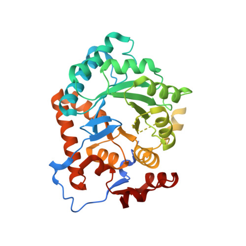
    Two types of isopentenyl diphosphate:dimethylallyl diphosphate isomerases (IDI) have been characterized at present. The long known IDI-1 is only dependent on divalent metals for activity, whereas IDI-2 requires a metal, FMN and NADPH. Here, we report the first structure of an IDI-2 from Bacillus subtilis at 1.9A resolution in the ligand-free form and of the FMN-bound form at 2.8A resolution. The enzyme is an octamer that forms a D4 symmetrical open, cage-like structure. The monomers of 45 kDa display a classical TIM barrel fold. FMN is bound only with very moderate affinity and is therefore completely lost during purification. However, the enzyme can be reconstituted in the crystals by soaking with FMN. Three glycine-rich sequence stretches that are characteristic for IDI-2 participate in FMN binding within the interior of the cage. Regions harboring strictly conserved residues that are implicated in substrate binding or catalysis remain largely disordered even in the presence of FMN.


  • Organizational Affiliation

    Abteilung für Strukturforschung, Max-Planck-Institut für Biochemie, Am Klopferspitz 18a, D-82152 Martinsried, Germany. steinbac@caltech.edu


Macromolecules
Find similar proteins by:  (by identity cutoff)  |  3D Structure
Entity ID: 1
MoleculeChains Sequence LengthOrganismDetailsImage
Isopentenyl-diphosphate delta-isomerase
A, B
349Bacillus subtilisMutation(s): 0 
EC: 5.3.3.2
UniProt
Find proteins for P50740 (Bacillus subtilis (strain 168))
Explore P50740 
Go to UniProtKB:  P50740
Entity Groups  
Sequence Clusters30% Identity50% Identity70% Identity90% Identity95% Identity100% Identity
UniProt GroupP50740
Sequence Annotations
Expand
  • Reference Sequence
Experimental Data & Validation

Experimental Data

  • Method: X-RAY DIFFRACTION
  • Resolution: 1.90 Å
  • R-Value Free: 0.221 
  • R-Value Work: 0.199 
  • R-Value Observed: 0.200 
  • Space Group: I 4
Unit Cell:
Length ( Å )Angle ( ˚ )
a = 115.833α = 90
b = 115.833β = 90
c = 141.52γ = 90
Software Package:
Software NamePurpose
DENZOdata reduction
SCALEPACKdata scaling
SHELXSphasing
CCP4model building
CNSrefinement
CCP4phasing

Structure Validation

View Full Validation Report


Entry History 

Deposition Data

Revision History  (Full details and data files)

  • Version 1.0: 2003-06-17
    Type: Initial release
  • Version 1.1: 2008-04-29
    Changes: Version format compliance
  • Version 1.2: 2011-07-13
    Changes: Derived calculations, Version format compliance
  • Version 1.3: 2024-03-13
    Changes: Data collection, Database references, Derived calculations