1OZJ

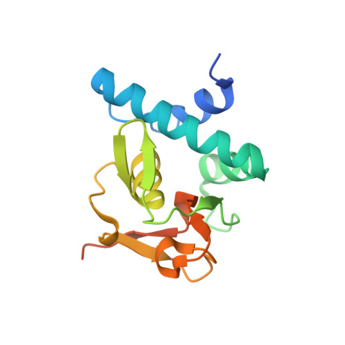
Crystal structure of Smad3-MH1 bound to DNA at 2.4 A resolution


Experimental Data Snapshot

  • Method: X-RAY DIFFRACTION
  • Resolution: 2.40 Å
  • R-Value Free: 0.270 
  • R-Value Work: 0.210 

wwPDB Validation   3D Report Full Report


This is version 1.4 of the entry. See complete history


Literature

Features of a Smad3 MH1-DNA complex. Roles of water and zinc in DNA binding.

Chai, J.Wu, J.-W.Yan, N.Massague, J.Pavletich, N.P.Shi, Y.

(2003) J Biol Chem 278: 20327-20331

  • DOI: https://doi.org/10.1074/jbc.C300134200
  • Primary Citation of Related Structures:  
    1OZJ

  • PubMed Abstract: 

    The Smad family of proteins mediates transforming growth factor-beta signaling from cell membrane to the nucleus. In the nucleus, Smads serve as transcription factors by directly binding to specific DNA sequences and regulating the expression of ligand-response genes. A previous structural analysis, at 2.8-A resolution, revealed a novel DNA-binding mode for the Smad MH1 domain but did not allow accurate assignment of the fines features of protein-DNA interactions. The crystal structure of a Smad3 MH1 domain bound to a palindromic DNA sequence, determined at 2.4-A resolution, reveals a surprisingly important role for water molecules. The asymmetric placement of the DNA-binding motif (a conserved 11-residue beta-hairpin) in the major groove of DNA is buttressed by seven well ordered water molecules. These water molecules make specific hydrogen bonds to the DNA bases, the DNA phosphate backbones, and several critical Smad3 residues. In addition, the MH1 domain is found to contain a bound zinc atom using four invariant residues among Smad proteins, three cysteines and one histidine. Removal of the zinc atom results in compromised DNA binding activity. These results define the Smad MH1 domain as a zinc-coordinating module that exhibits unique DNA binding properties.


  • Organizational Affiliation

    Department of Molecular Biology, Princeton University, Lewis Thomas Laboratory, New Jersey 08544, USA.


Macromolecules

Find similar proteins by:  (by identity cutoff)  |  3D Structure
Entity ID: 3
MoleculeChains Sequence LengthOrganismDetailsImage
SMAD 3C [auth A],
D [auth B]
144Homo sapiensMutation(s): 0 
Gene Names: MADH3 OR SMAD3 OR MAD3
UniProt & NIH Common Fund Data Resources
Find proteins for P84022 (Homo sapiens)
Explore P84022 
Go to UniProtKB:  P84022
PHAROS:  P84022
GTEx:  ENSG00000166949 
Entity Groups  
Sequence Clusters30% Identity50% Identity70% Identity90% Identity95% Identity100% Identity
UniProt GroupP84022
Sequence Annotations
Expand
  • Reference Sequence

Find similar nucleic acids by:  Sequence   |   3D Structure  

Entity ID: 1
MoleculeChains LengthOrganismImage
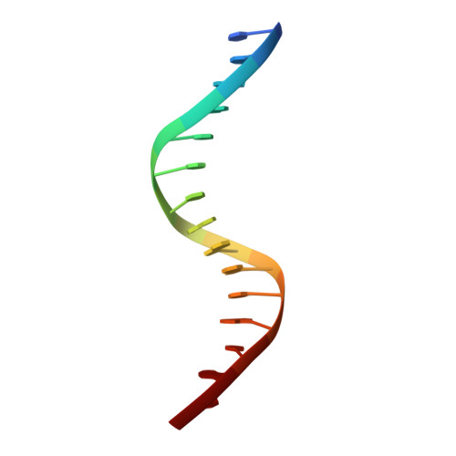
Smad binding elementA [auth C]15N/A
Sequence Annotations
Expand
  • Reference Sequence

Find similar nucleic acids by:  Sequence   |   3D Structure  

Entity ID: 2
MoleculeChains LengthOrganismImage
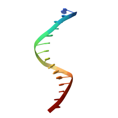
Smad binding elementB [auth D]15N/A
Sequence Annotations
Expand
  • Reference Sequence
Small Molecules
Ligands 1 Unique
IDChains Name / Formula / InChI Key2D Diagram3D Interactions
ZN
Query on ZN

Download Ideal Coordinates CCD File 
E [auth A],
F [auth B]
ZINC ION
Zn
PTFCDOFLOPIGGS-UHFFFAOYSA-N
Experimental Data & Validation

Experimental Data

  • Method: X-RAY DIFFRACTION
  • Resolution: 2.40 Å
  • R-Value Free: 0.270 
  • R-Value Work: 0.210 
  • Space Group: P 1 21 1
Unit Cell:
Length ( Å )Angle ( ˚ )
a = 45.6α = 90
b = 60.4β = 102
c = 71.6γ = 90
Software Package:
Software NamePurpose
DENZOdata reduction
SCALEPACKdata scaling
AMoREphasing
CNSrefinement

Structure Validation

View Full Validation Report


Entry History 

Deposition Data

Revision History  (Full details and data files)

  • Version 1.0: 2004-03-23
    Type: Initial release
  • Version 1.1: 2008-04-29
    Changes: Version format compliance
  • Version 1.2: 2011-07-13
    Changes: Version format compliance
  • Version 1.3: 2018-04-04
    Changes: Data collection
  • Version 1.4: 2024-02-14
    Changes: Data collection, Database references, Derived calculations