Probing the roles of key residues in the unique regulatory NADH binding site of type II citrate synthase of Escherichia coli.
Stokell, D.J., Donald, L.J., Maurus, R., Nguyen, N.T., Sadler, G., Choudhary, K., Hultin, P.G., Brayer, G.D., Duckworth, H.W.(2003) J Biol Chem 278: 35435-35443
- PubMed: 12824188
- DOI: https://doi.org/10.1074/jbc.M302786200
- Primary Citation of Related Structures:
1OWB, 1OWC - PubMed Abstract:
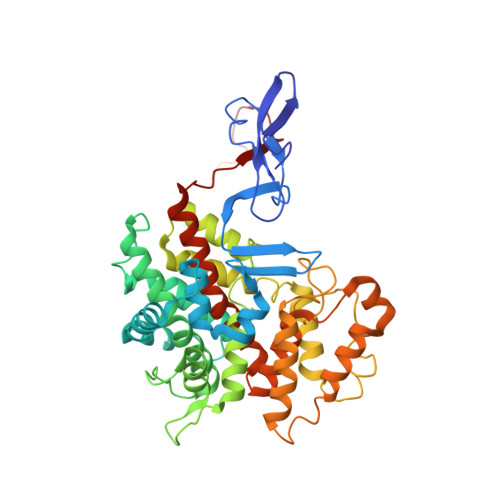
The citrate synthase of Escherichia coli is an example of a Type II citrate synthase, a hexamer that is subject to allosteric inhibition by NADH. In previous crystallographic work, we defined the NADH binding sites, identifying nine amino acids whose side chains were proposed to make hydrogen bonds with the NADH molecule. Here, we describe the functional properties of nine sequence variants, in which these have been replaced by nonbonding residues. All of the variants show some changes in NADH binding and inhibition and small but significant changes in kinetic parameters for catalysis. In three cases, Y145A, R163L, and K167A, NADH inhibition has become extremely weak. We have used nanospray/time-of-flight mass spectrometry, under non-denaturing conditions, to show that two of these, R163L and K167A, do not form hexamers in response to NADH binding, unlike the wild type enzyme. One variant, R109L, shows tighter NADH binding. We have crystallized this variant and determined its structure, with and without bound NADH. Unexpectedly, the greatest structural changes in the R109L variant are in two regions outside the NADH binding site, both of which, in wild type citrate synthase, have unusually high mobilities as measured by crystallographic thermal factors. In the R109L variant, both regions (residues 260 -311 and 316-342) are much less mobile and have rearranged significantly. We argue that these two regions are elements in the path of communication between the NADH binding sites and the active sites and are centrally involved in the regulatory conformational change in E. coli citrate synthase.
Organizational Affiliation:
Department of Chemistry, University of Manitoba, Winnipeg, Manitoba R3T 2N2, Canada.