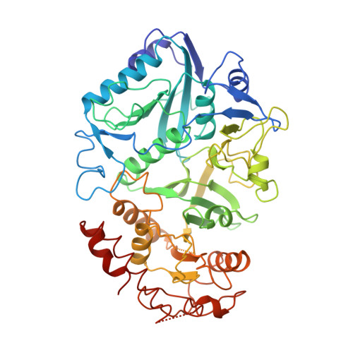
Crystal structure of Escherichia coli phosphoenolpyruvate carboxykinase: a new structural family with the P-loop nucleoside triphosphate hydrolase fold.
Matte, A., Goldie, H., Sweet, R.M., Delbaere, L.T.(1996) J Mol Biol 256: 126-143
- PubMed: 8609605
- DOI: https://doi.org/10.1006/jmbi.1996.0072
- Primary Citation of Related Structures:
1OEN - PubMed Abstract:
The crystal structure of ATP-dependent phosphoenolpyruvate carboxykinase (ATP-oxaloacetate carboxy-lyase, (transphosphorylating), E.C. 4.1.1.49; PCK) from Escherichia coli strain K12 has been determined using a combination of multiple isomorphous replacement, density modification, and partial model phase combination, and refined to a conventional R-index of 0.204 (Rfree = 0.244) at 1.9 A resolution. Each PCK molecule consists of a 275 residue N-terminal domain and 265 residue C-terminal or mononucleotide-binding domain, with the active site postulated to be within a cleft between the two domains. PCK is an open-faced, mixed alpha/beta protein, with each domain having an alpha/beta folding topology as found in several other mononucleoside-binding enzymes. The putative phosphate-binding site of ATP adopts the P-loop motif common to many ATP and GTP-binding proteins, and is similar in structure to that found within adenylate kinase. However, the beta-sheet topology within the mononucleotide-binding fold of PCK differs from all other families within the P-loop containing nucleoside triphosphate hydrolase superfamily, therefore suggesting it represents the first member in a new family of such proteins. The mononucleotide-binding domain is also different in structure compared to the classical mononucleotide-binding fold (CMBF) common to adenylate kinase, p21ras, and elongation factor-Tu. Several amino acid residues, including R65, K212, K213, H232, K254, D269, K288 and R333 appear to make up the active site of the enzyme, and are found to be absolutely conserved among known members of the ATP-dependent PCK family. A cysteine residue is located near the active-site, as has been suggested for other PCKs, although in the E. coli enzyme C233 is buried and so is most likely not involved in substrate binding or catalysis. Two binding sites of the calcium-analog TB3+ have been determined, one within the active site coordinating to the side-chain of D269, and the other within the C-terminal domain coordinating to the side-chains of E508 and E511.
Organizational Affiliation:
Department of Biochemistry, University of Saskatchewan Saskatoon, Canada.


