1OD6

The Crystal Structure of Phosphopantetheine adenylyltransferase from Thermus Thermophilus in complex with 4'-phosphopantetheine


Experimental Data Snapshot

  • Method: X-RAY DIFFRACTION
  • Resolution: 1.50 Å
  • R-Value Free: 0.201 
  • R-Value Work: 0.196 
  • R-Value Observed: 0.196 

wwPDB Validation   3D Report Full Report


Ligand Structure Quality Assessment 


This is version 1.2 of the entry. See complete history


Literature

Structure and Implications for the Thermal Stability of Phosphopantetheine Adenylyltransferase from Thermus Thermophilus.

Takahashi, H.Inagaki, E.Fujimoto, Y.Kuroishi, C.Nodake, Y.Nakamura, Y.Arisaka, F.Yutani, K.Kuramitsu, S.Yokoyama, S.Yamamoto, M.Miyano, M.Tahirov, T.H.

(2004) Acta Crystallogr D Biol Crystallogr 60: 97

  • DOI: https://doi.org/10.1107/s0907444903025319
  • Primary Citation of Related Structures:  
    1OD6

  • PubMed Abstract: 

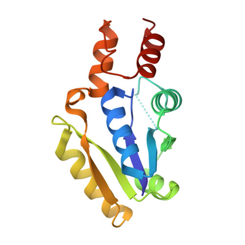
    Phosphopantetheine adenylyltransferase (PPAT) is an essential enzyme in bacteria that catalyzes the rate-limiting step in coenzyme A (CoA) biosynthesis by transferring an adenylyl group from ATP to 4'-phosphopantetheine (Ppant), yielding 3'-dephospho-CoA (dPCoA). The crystal structure of PPAT from Thermus thermophilus HB8 (Tt PPAT) complexed with Ppant has been determined by the molecular-replacement method at 1.5 A resolution. The overall fold of the enzyme is almost the same as that of Escherichia coli PPAT, a hexamer having point group 32. The asymmetric unit of Tt PPAT contains a monomer and the crystallographic triad and dyad coincide with the threefold and twofold axes of the hexamer, respectively. Most of the important atoms surrounding the active site in E. coli PPAT are conserved in Tt PPAT, indicating similarities in their substrate binding and enzymatic reaction. The notable difference between E. coli PPAT and Tt PPAT is the simultaneous substrate recognition by all six subunits of Tt PPAT compared with substrate recognition by only three subunits in E. coli PPAT. Comparative analysis also revealed that the higher stability of Tt PPAT arises from stabilization of each subunit by hydrophobic effects, hydrogen bonds and entropic effects.


  • Organizational Affiliation

    Highthroughput Factory, RIKEN Harima Institute, 1-1-1 Kouto, Mikazuki-cho, Sayo-gun, Hyogo 679-5148, Japan.


Macromolecules
Find similar proteins by:  (by identity cutoff)  |  3D Structure
Entity ID: 1
MoleculeChains Sequence LengthOrganismDetailsImage
PHOSPHOPANTETHEINE ADENYLYLTRANSFERASE160Thermus thermophilus HB8Mutation(s): 0 
EC: 2.7.7.3
UniProt
Find proteins for Q5SJS9 (Thermus thermophilus (strain ATCC 27634 / DSM 579 / HB8))
Explore Q5SJS9 
Go to UniProtKB:  Q5SJS9
Entity Groups  
Sequence Clusters30% Identity50% Identity70% Identity90% Identity95% Identity100% Identity
UniProt GroupQ5SJS9
Sequence Annotations
Expand
  • Reference Sequence
Experimental Data & Validation

Experimental Data

  • Method: X-RAY DIFFRACTION
  • Resolution: 1.50 Å
  • R-Value Free: 0.201 
  • R-Value Work: 0.196 
  • R-Value Observed: 0.196 
  • Space Group: H 3 2
Unit Cell:
Length ( Å )Angle ( ˚ )
a = 115.715α = 90
b = 115.715β = 90
c = 115.661γ = 120
Software Package:
Software NamePurpose
CNSrefinement
DENZOdata reduction
SCALEPACKdata scaling
CNSphasing

Structure Validation

View Full Validation Report


Ligand Structure Quality Assessment 


Entry History 

Deposition Data

Revision History  (Full details and data files)

  • Version 1.0: 2003-03-13
    Type: Initial release
  • Version 1.1: 2015-12-16
    Changes: Database references, Derived calculations, Non-polymer description, Other, Refinement description, Structure summary, Version format compliance
  • Version 1.2: 2023-12-13
    Changes: Data collection, Database references, Derived calculations, Other, Refinement description