De Novo Phasing of Two Crystal Forms of Tryparedoxin II Using the Anomalous Scattering from S Atoms: A Combination of Small Signal and Medium Resolution Reveals This to be a General Tool for Solving Protein Crystal Structures
Micossi, E., Hunter, W.N., Leonard, G.A.(2002) Acta Crystallogr D Biol Crystallogr 58: 21
- PubMed: 11752776
- DOI: https://doi.org/10.1107/s0907444901016808
- Primary Citation of Related Structures:
1O6J, 1O81 - PubMed Abstract:
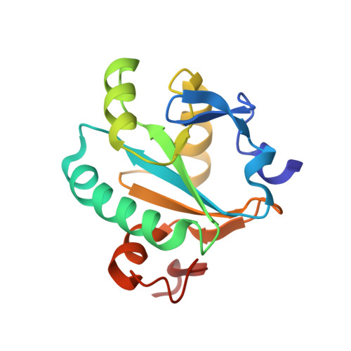
The de novo phasing of the structures of two crystal forms of tryparedoxin II from Crithidia fasciculata has been carried out using single-wavelength anomalous diffraction techniques exploiting only the small anomalous signal from the S atoms intrinsic to the native protein. Data were collected at 1.77 A wavelength, where the Bijvoet ratio is approximately 1.2%. Data collected to d(min) = 2.5 A from a crystal of form I, which has a diffraction limit of d(min) = 1.5 A and a solvent content of approximately 46%, produced readily interpretable electron-density maps. When these phases were extended to the resolution limit of the crystals, almost the entire model could be traced automatically. Crystals of form II have a much higher solvent content, approximately 72%, and a much lower diffraction limit than form I and at 1.77 A wavelength yielded data only to d(min) = 2.7 A. Despite the medium resolution of the data for this crystal form, it was possible both to determine the heavy-atom partial structure and then use it to produce, still at d(min) = 2.7 A, an excellent quality interpretable electron-density map. This was then improved by phase extension to the d(min) = 2.35 A diffraction limits of a different crystal for which data were collected on a more intense beamline. The success of this latter structure solution markedly increases the potential use in macromolecular crystal structure determination of the anomalous signal available from S atoms that occur naturally in proteins and, as is discussed, has significant implications for structure determination in the high-throughput era.
Organizational Affiliation:
Macromolecular Crystallography, European Synchrotron Radiation Facility, BP 220, F-38043 Grenoble CEDEX, France.