1NMA

N9 NEURAMINIDASE COMPLEXES WITH ANTIBODIES NC41 AND NC10: EMPIRICAL FREE-ENERGY CALCULATIONS CAPTURE SPECIFICITY TRENDS OBSERVED WITH MUTANT BINDING DATA


Experimental Data Snapshot

  • Method: X-RAY DIFFRACTION
  • Resolution: 3.00 Å
  • R-Value Work: 0.200 
  • R-Value Observed: 0.200 

wwPDB Validation   3D Report Full Report


This is version 2.0 of the entry. See complete history


Literature

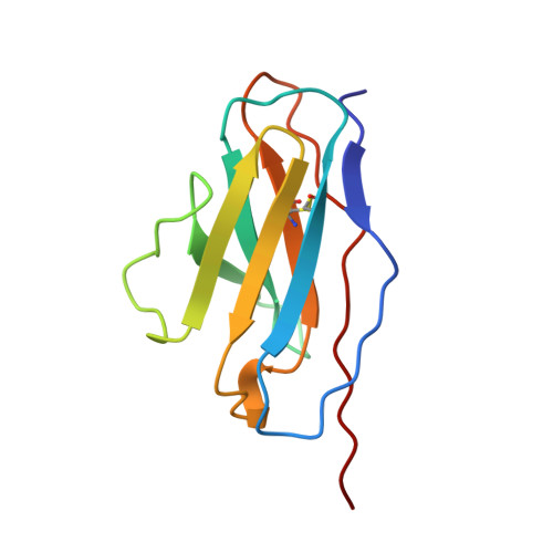
N9 neuraminidase complexes with antibodies NC41 and NC10: empirical free energy calculations capture specificity trends observed with mutant binding data.

Tulip, W.R.Harley, V.R.Webster, R.G.Novotny, J.

(1994) Biochemistry 33: 7986-7997

  • DOI: https://doi.org/10.1021/bi00192a002
  • Primary Citation of Related Structures:  
    1NMA

  • PubMed Abstract: 

    X-ray crystallographic coordinates of influenza virus N9 neuraminidase complexed with monoclonal antibodies NC41 and NC10 [Tulip et al. (1992) J. Mol. Biol. 227, 122-148] served as a starting point for calculations aimed at estimating free energy changes (delta G) of complex formation between the two antibodies and the neuraminidase. Using an empirical function incorporating hydrophobic, electrostatic, and conformational entropy effects, we estimated contributions individual neuraminidase residues make to complex formation (delta G(residue)) and compared the calculated values to experimentally measured differences in antibody binding between the wild-type and mutated neuraminidases [Nuss et al. (1993) Proteins 15, 121-132; calculations done without prior knowledge of the experimental data]. A good correspondence was found between the calculated delta G(residue) values and the mutant binding data in that side chains with large calculated delta G contributions (delta G(residue) < -1 kcal/mol) lie at sites of mutation which cause a marked reduction in antibody binding, and side chains for which delta G(residue) > -1 kcal/mol are sites at which a mutation does not have a marked effect on binding. Because most of the delta G(residue) < -1 kcal/mol side chains also make hydrogen bonds/salt bridges with the antibody, the correspondence of the effect of antibody binding with these electrostatic interactions (18 out of 27 for NC41 and, tentatively, 5 out of 7 for NC10) is about as good as that with predicted energetic residues. All the delta G(residue) < -1 kcal/mol neuraminidase side chains cluster around the most protruding surface regions and are thus spread over different epitope segments. Surprisingly, different residues were found to make the most critical contributions to the NC41 and NC10 complex stabilities despite the fact that the NC41 and NC10 antigenic epitopes overlap, having approximately 70% of surface residues in common. It is thus possible, for two different antibodies, to recognize the same protein surface in strikingly different ways. As only a fraction of the neuraminidase residues appear to make large contributions to antibody binding, the results also support the hypothesis of a "functional" epitope in antigen-antibody interactions. Positive trends between both backbone rigidity and residue accessibility in the complexed state, and contributions of these residues to binding, were also observed for the NC41 complex.


  • Organizational Affiliation

    CSIRO Division of Biomolecular Engineering, Parkville, Victoria, Australia.


Macromolecules
Find similar proteins by:  (by identity cutoff)  |  3D Structure
Entity ID: 1
MoleculeChains Sequence LengthOrganismDetailsImage
N9 NEURAMINIDASEA [auth N]388Influenza A virusMutation(s): 0 
EC: 3.2.1.18
UniProt
Find proteins for P05803 (Influenza A virus (strain A/Whale/Maine/1/1984 H13N9))
Explore P05803 
Go to UniProtKB:  P05803
Entity Groups  
Sequence Clusters30% Identity50% Identity70% Identity90% Identity95% Identity100% Identity
UniProt GroupP05803
Sequence Annotations
Expand
  • Reference Sequence
Find similar proteins by:  (by identity cutoff)  |  3D Structure
Entity ID: 2
MoleculeChains Sequence LengthOrganismDetailsImage
FAB NC10B [auth L]109Mus musculusMutation(s): 0 
Entity Groups  
Sequence Clusters30% Identity50% Identity70% Identity90% Identity95% Identity100% Identity
Sequence Annotations
Expand
  • Reference Sequence
Find similar proteins by:  (by identity cutoff)  |  3D Structure
Entity ID: 3
MoleculeChains Sequence LengthOrganismDetailsImage
FAB NC10C [auth H]122Mus musculusMutation(s): 0 
Entity Groups  
Sequence Clusters30% Identity50% Identity70% Identity90% Identity95% Identity100% Identity
Sequence Annotations
Expand
  • Reference Sequence
Oligosaccharides

Help

Entity ID: 4
MoleculeChains Length2D Diagram Glycosylation3D Interactions
alpha-D-mannopyranose-(1-2)-alpha-D-mannopyranose-(1-2)-alpha-D-mannopyranose-(1-3)-beta-D-mannopyranose-(1-4)-2-acetamido-2-deoxy-beta-D-glucopyranose-(1-4)-2-acetamido-2-deoxy-beta-D-glucopyranoseD [auth A]6N-Glycosylation
Glycosylation Resources
GlyTouCan:  G01760ZU
GlyCosmos:  G01760ZU
GlyGen:  G01760ZU
Small Molecules
Ligands 1 Unique
IDChains Name / Formula / InChI Key2D Diagram3D Interactions
NAG
Query on NAG

Download Ideal Coordinates CCD File 
E [auth N]2-acetamido-2-deoxy-beta-D-glucopyranose
C8 H15 N O6
OVRNDRQMDRJTHS-FMDGEEDCSA-N
Experimental Data & Validation

Experimental Data

  • Method: X-RAY DIFFRACTION
  • Resolution: 3.00 Å
  • R-Value Work: 0.200 
  • R-Value Observed: 0.200 
  • Space Group: I 4 2 2
Unit Cell:
Length ( Å )Angle ( ˚ )
a = 171.5α = 90
b = 171.5β = 90
c = 160.2γ = 90
Software Package:
Software NamePurpose
X-PLORmodel building
X-PLORrefinement
X-PLORphasing

Structure Validation

View Full Validation Report


Entry History 

Deposition Data

Revision History  (Full details and data files)

  • Version 1.0: 1995-09-15
    Type: Initial release
  • Version 1.1: 2008-03-24
    Changes: Version format compliance
  • Version 1.2: 2011-07-13
    Changes: Non-polymer description, Version format compliance
  • Version 2.0: 2020-07-29
    Type: Remediation
    Reason: Carbohydrate remediation
    Changes: Atomic model, Data collection, Database references, Derived calculations, Other, Structure summary