Structure and function of S-adenosylmethionine synthetase: crystal structures of S-adenosylmethionine synthetase with ADP, BrADP, and PPi at 28 angstroms resolution.
Takusagawa, F., Kamitori, S., Markham, G.D.(1996) Biochemistry 35: 2586-2596
- PubMed: 8611562
- DOI: https://doi.org/10.1021/bi952604z
- Primary Citation of Related Structures:
1MXA, 1MXB, 1MXC - PubMed Abstract:
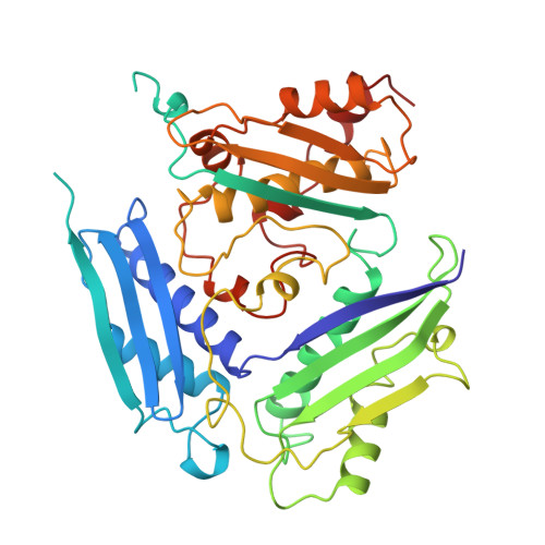
S-Adenosylmethionine synthetase (MAT,ATP:L-methionine S-adenosltransferase, EC 2.5.1.6) plays a central metabolic role in all organisms. MAT catalyzes the two-step reaction which synthesizes S-adenosylmethionine (AdoMet), pyrophosphate (PPi), and orthophosphate (Pi) from ATP and L-methionine. AdoMet is the primary methyl group donor in biological systems. The first crystal structure of MAT from Escherichia coli has recently been determined [Takusagawa et al. (1995) J. Biol. Chem. 271, 136-147]. In order to elucidate the active site and possible catalytic reaction mechanism, the MAT structures in the crystals grown with the substrate ATP (and BrATP) and the product PPi have been determined (space group P6(2)22; unit cell a = b = 128.9 Angstroms, c= 139.8 Angstroms, resolution limit 2.8 Angstroms; R O.19; Rfree 0.26). The enzyme consists of four identical subunits; two subunits form a spherical dimer, and pairs of these tightly bound dimers form a tetrameric enzyme. Each dimer has two active sites which are located between the subunits. Each subunit consists of three domains related to each other by a pseudo 3-fold symmetry. The crystal structures showed that the ATP molecules were hydrolyzed to ADP and Pi by the enzyme. Those products were found at the active site along with the essential metal ions (K+ and Mg2+). This rather unexpected finding was first confirmed by the structure of the complex with PPi and later by an HPLC analysis. The enzyme hydrolyzed ATP to ADP and Pi in 72 h under the same conditions as the crystallization of the enzyme. In the active site, the diphosphate moiety of ADP and Pi interacts extensively with amino acid residues from the two subunits of the enzyme, whereas the adenine and ribose moieties have little interaction with the enzyme. The enzyme structure is little changed upon binding ADP. All amino acid residues involved in the active site are found to be conserved in the 14 reported sequences of MAT from a wide range of organisms. Thus the structure determined in this study can be utilized as a model for other members of the MAT family. On the basis of the crystal structures, the catalytic reaction mechanisms of AdoMet formation and hydrolysis of tripolyphosphate are proposed.
Organizational Affiliation:
Department of Chemistry, University of Kansas, Lawrence, Kansas 66045-0046, USA.