1MOV

Crystal structure of Coral protein mutant


Experimental Data Snapshot

  • Method: X-RAY DIFFRACTION
  • Resolution: 2.40 Å
  • R-Value Free: 0.249 
  • R-Value Work: 0.225 
  • R-Value Observed: 0.225 

wwPDB Validation   3D Report Full Report


This is version 2.0 of the entry. See complete history


Literature

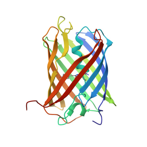
The 2.2 a crystal structure of a pocilloporin pigment reveals a nonplanar chromophore conformation.

Prescott, M.Ling, M.Beddoe, T.Oakley, A.J.Dove, S.Hoegh-Guldberg, O.Devenish, R.J.Rossjohn, J.

(2003) Structure 11: 275-284

  • DOI: https://doi.org/10.1016/s0969-2126(03)00028-5
  • Primary Citation of Related Structures:  
    1MOU, 1MOV

  • PubMed Abstract: 

    Reef-building corals contain host pigments, termed pocilloporins, that function to regulate the light environment of their resident microalgae by acting as a photoprotectant in excessive sunlight. We have determined the crystal structure of an intensely blue, nonfluorescent pocilloporin to 2.2 A resolution and a genetically engineered fluorescent variant to 2.4 A resolution. The pocilloporin chromophore structure adopts a markedly different conformation in comparison with the DsRed chromophore, despite the chromophore sequences (Gln-Tyr-Gly) being identical; the tyrosine ring of the pocilloporin chromophore is noncoplanar and in the trans configuration. Furthermore, the fluorescent variant adopted a noncoplanar chromophore conformation. The data presented here demonstrates that the conformation of the chromophore is highly dependent on its immediate environment.


  • Organizational Affiliation

    Department of Biochemistry and Molecular Biology, School of Biomedical Sciences, Monash University, Clayton, 3800, Victoria, Australia. mark.prescott@med.monash.au


Macromolecules
Find similar proteins by:  (by identity cutoff)  |  3D Structure
Entity ID: 1
MoleculeChains Sequence LengthOrganismDetailsImage
GFP-like non-fluorescent chromoprotein219Montipora efflorescensMutation(s): 2 
UniProt
Find proteins for P83690 (Montipora efflorescens)
Explore P83690 
Go to UniProtKB:  P83690
Entity Groups  
Sequence Clusters30% Identity50% Identity70% Identity90% Identity95% Identity100% Identity
UniProt GroupP83690
Sequence Annotations
Expand
  • Reference Sequence
Experimental Data & Validation

Experimental Data

  • Method: X-RAY DIFFRACTION
  • Resolution: 2.40 Å
  • R-Value Free: 0.249 
  • R-Value Work: 0.225 
  • R-Value Observed: 0.225 
  • Space Group: I 41 2 2
Unit Cell:
Length ( Å )Angle ( ˚ )
a = 131.391α = 90
b = 131.391β = 90
c = 150.67γ = 90
Software Package:
Software NamePurpose
AMoREphasing
CNSrefinement

Structure Validation

View Full Validation Report


Entry History 

Deposition Data

Revision History  (Full details and data files)

  • Version 1.0: 2003-04-08
    Type: Initial release
  • Version 1.1: 2008-04-28
    Changes: Version format compliance
  • Version 1.2: 2011-07-13
    Changes: Derived calculations, Version format compliance
  • Version 1.3: 2017-10-11
    Changes: Refinement description
  • Version 1.4: 2021-11-10
    Changes: Database references, Derived calculations
  • Version 1.5: 2023-10-25
    Changes: Data collection, Refinement description
  • Version 2.0: 2023-11-15
    Changes: Atomic model, Data collection, Derived calculations