1MKW

THE CO-CRYSTAL STRUCTURE OF UNLIGANDED BOVINE ALPHA-THROMBIN AND PRETHROMBIN-2: MOVEMENT OF THE YPPW SEGMENT AND ACTIVE SITE RESIDUES UPON LIGAND BINDING


Experimental Data Snapshot

  • Method: X-RAY DIFFRACTION
  • Resolution: 2.30 Å
  • R-Value Free: 0.282 
  • R-Value Work: 0.190 
  • R-Value Observed: 0.190 

wwPDB Validation   3D Report Full Report


This is version 1.2 of the entry. See complete history


Literature

The co-crystal structure of unliganded bovine alpha-thrombin and prethrombin-2: movement of the Tyr-Pro-Pro-Trp segment and active site residues upon ligand binding.

Malkowski, M.G.Martin, P.D.Guzik, J.C.Edwards, B.F.

(1997) Protein Sci 6: 1438-1448

  • DOI: https://doi.org/10.1002/pro.5560060708
  • Primary Citation of Related Structures:  
    1MKW, 1MKX

  • PubMed Abstract: 

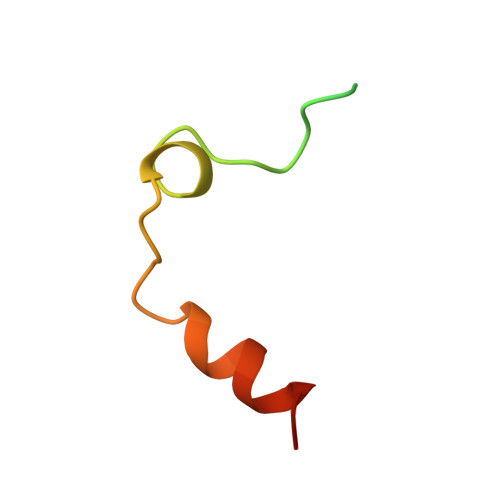
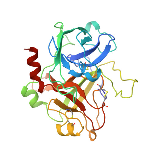
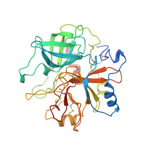
    Unliganded bovine alpha-thrombin and prethrombin-2 have been co-crystallized, in space group P21212, using either ammonium sulfate or polyethylene glycol 2000 (PEG2K), and their structures determined at 2.2 A and 2.3 A, respectively. Initial phases were determined by molecular replacement and refined using XPLOR to final R factors of 0.187 (Rfree = 0.255) and 0.190 (Rfree = 0.282) for the salt and PEG2K models, respectively. The apo-enzyme form of bovine alpha-thrombin shows dramatic shifts in placement for the Tyr-Pro-Pro-Trp segment, for Glu-192, and for the catalytic residues His-57 and Ser-195, when compared to 4 thrombin complexes representing different states of catalysis, namely (1) the Michaelis complex (residues 7-19 of fibrinogen A alpha with a non-cleavable scissile bond), (2) enzyme-inhibitor complex (D-Phe-Pro-Arg chloromethylketone), (3) enzyme product complex (residues 7-16 of fibrinopeptide A), and (4) the exosite complex (residues 53-64 of hirudin). The structures of bovine and human prethrombin-2 are generally similar to one another (RMS deviation of 0.68 A) but differ significantly in the Arg-15/Ile-16 cleavage region and in the three activation domains, which are disordered in bovine prethrombin-2, analogous to that seen for trypsinogen.


  • Organizational Affiliation

    Department of Biochemistry and Molecular Biology, Wayne State University, Detroit, Michigan 48201, USA.


Macromolecules
Find similar proteins by:  (by identity cutoff)  |  3D Structure
Entity ID: 1
MoleculeChains Sequence LengthOrganismDetailsImage
ALPHA-THROMBINA [auth L]49Bos taurusMutation(s): 0 
EC: 3.4.21.5
UniProt
Find proteins for P00735 (Bos taurus)
Explore P00735 
Go to UniProtKB:  P00735
Entity Groups  
Sequence Clusters30% Identity50% Identity70% Identity90% Identity95% Identity100% Identity
UniProt GroupP00735
Sequence Annotations
Expand
  • Reference Sequence
Find similar proteins by:  (by identity cutoff)  |  3D Structure
Entity ID: 2
MoleculeChains Sequence LengthOrganismDetailsImage
ALPHA-THROMBINB [auth H]259Bos taurusMutation(s): 0 
EC: 3.4.21.5
UniProt
Find proteins for P00735 (Bos taurus)
Explore P00735 
Go to UniProtKB:  P00735
Entity Groups  
Sequence Clusters30% Identity50% Identity70% Identity90% Identity95% Identity100% Identity
UniProt GroupP00735
Sequence Annotations
Expand
  • Reference Sequence
Find similar proteins by:  (by identity cutoff)  |  3D Structure
Entity ID: 3
MoleculeChains Sequence LengthOrganismDetailsImage
PRETHROMBIN-2C [auth K]308Bos taurusMutation(s): 0 
EC: 3.4.21.5
UniProt
Find proteins for P00735 (Bos taurus)
Explore P00735 
Go to UniProtKB:  P00735
Entity Groups  
Sequence Clusters30% Identity50% Identity70% Identity90% Identity95% Identity100% Identity
UniProt GroupP00735
Sequence Annotations
Expand
  • Reference Sequence
Experimental Data & Validation

Experimental Data

  • Method: X-RAY DIFFRACTION
  • Resolution: 2.30 Å
  • R-Value Free: 0.282 
  • R-Value Work: 0.190 
  • R-Value Observed: 0.190 
  • Space Group: P 21 21 2
Unit Cell:
Length ( Å )Angle ( ˚ )
a = 87.58α = 90
b = 88.55β = 90
c = 101.03γ = 90
Software Package:
Software NamePurpose
XENGENdata collection
XENGENdata reduction
X-PLORmodel building
X-PLORrefinement
XENGENdata scaling
X-PLORphasing

Structure Validation

View Full Validation Report


Entry History 

Deposition Data

Revision History  (Full details and data files)

  • Version 1.0: 1997-07-07
    Type: Initial release
  • Version 1.1: 2008-03-24
    Changes: Version format compliance
  • Version 1.2: 2011-07-13
    Changes: Version format compliance