1MGV

Crystal Structure of the R391A Mutant of 7,8-Diaminopelargonic Acid Synthase


Experimental Data Snapshot

  • Method: X-RAY DIFFRACTION
  • Resolution: 2.10 Å
  • R-Value Free: 0.233 
  • R-Value Work: 0.201 
  • R-Value Observed: 0.203 

wwPDB Validation   3D Report Full Report


Ligand Structure Quality Assessment 


This is version 1.5 of the entry. See complete history


Literature

The Dual-Specific Active Site of 7,8-Diaminopelargonic Acid Synthase and the Effect of the R391A Mutation

Eliot, A.C.Sandmark, J.Schneider, G.Kirsch, J.F.

(2002) Biochemistry 41: 12582-12589

  • DOI: https://doi.org/10.1021/bi026339a
  • Primary Citation of Related Structures:  
    1MGV

  • PubMed Abstract: 

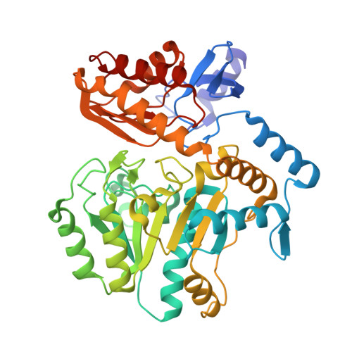
    7,8-diaminopelargonic acid (DAPA) synthase (EC 2.6.1.62) is a pyridoxal phosphate (PLP)-dependent transaminase that catalyzes the transfer of the alpha-amino group from S-adenosyl-L-methionine (SAM) to 7-keto-8-aminopelargonic acid (KAPA) to form DAPA in the antepenultimate step in the biosynthesis of biotin. The wild-type enzyme has a steady-state kcat value of 0.013 s(-1), and the K(m) values for SAM and KAPA are 150 and <2 microM, respectively. The k(max) and apparent K(m) values for the half-reaction of the PLP form of the enzyme with SAM are 0.016 s(-1) and 300 microM, respectively, while those for the reaction with DAPA are 0.79 s(-1) and 1 microM. The R391A mutant enzyme exhibits near wild-type kinetic parameters in the reaction with SAM, while the apparent K(m) for DAPA is increased 180-fold. The 2.1 A crystal structure of the R391A mutant enzyme shows that the mutation does not significantly alter the structure. These results indicate that the conserved arginine residue is not required for binding the alpha-amino acid SAM, but it is important for recognition of DAPA.


  • Organizational Affiliation

    Department of Chemistry, University of California-Berkeley, Berkeley, California 94720-3206, USA.


Macromolecules
Find similar proteins by:  (by identity cutoff)  |  3D Structure
Entity ID: 1
MoleculeChains Sequence LengthOrganismDetailsImage
7,8-diamino-pelargonic acid aminotransferase
A, B
429Escherichia coliMutation(s): 1 
Gene Names: bioA
EC: 2.6.1.62
UniProt
Find proteins for P12995 (Escherichia coli (strain K12))
Explore P12995 
Go to UniProtKB:  P12995
Entity Groups  
Sequence Clusters30% Identity50% Identity70% Identity90% Identity95% Identity100% Identity
UniProt GroupP12995
Sequence Annotations
Expand
  • Reference Sequence
Small Molecules
Ligands 3 Unique
IDChains Name / Formula / InChI Key2D Diagram3D Interactions
PLP
Query on PLP

Download Ideal Coordinates CCD File 
D [auth A],
G [auth B]
PYRIDOXAL-5'-PHOSPHATE
C8 H10 N O6 P
NGVDGCNFYWLIFO-UHFFFAOYSA-N
IPA
Query on IPA

Download Ideal Coordinates CCD File 
E [auth A],
H [auth B],
I [auth B],
J [auth B]
ISOPROPYL ALCOHOL
C3 H8 O
KFZMGEQAYNKOFK-UHFFFAOYSA-N
NA
Query on NA

Download Ideal Coordinates CCD File 
C [auth A],
F [auth B]
SODIUM ION
Na
FKNQFGJONOIPTF-UHFFFAOYSA-N
Experimental Data & Validation

Experimental Data

  • Method: X-RAY DIFFRACTION
  • Resolution: 2.10 Å
  • R-Value Free: 0.233 
  • R-Value Work: 0.201 
  • R-Value Observed: 0.203 
  • Space Group: C 1 2 1
Unit Cell:
Length ( Å )Angle ( ˚ )
a = 127.88α = 90
b = 55.899β = 110.06
c = 116.237γ = 90
Software Package:
Software NamePurpose
MOSFLMdata reduction
SCALAdata scaling
AMoREphasing
REFMACrefinement
CCP4data scaling

Structure Validation

View Full Validation Report


Ligand Structure Quality Assessment 


Entry History 

Deposition Data

Revision History  (Full details and data files)

  • Version 1.0: 2002-11-27
    Type: Initial release
  • Version 1.1: 2008-04-28
    Changes: Version format compliance
  • Version 1.2: 2011-07-13
    Changes: Non-polymer description, Version format compliance
  • Version 1.3: 2018-03-07
    Changes: Advisory, Data collection
  • Version 1.4: 2021-10-27
    Changes: Advisory, Database references, Derived calculations
  • Version 1.5: 2024-04-03
    Changes: Data collection, Refinement description