1MEG

CRYSTAL STRUCTURE OF A CARICAIN D158E MUTANT IN COMPLEX WITH E-64


Experimental Data Snapshot

  • Method: X-RAY DIFFRACTION
  • Resolution: 2.00 Å
  • R-Value Work: 0.193 
  • R-Value Observed: 0.193 

wwPDB Validation   3D Report Full Report


Ligand Structure Quality Assessment 


This is version 1.3 of the entry. See complete history


Literature

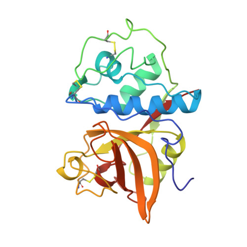
Crystal structure of a caricain D158E mutant in complex with E-64.

Katerelos, N.A.Taylor, M.A.Scott, M.Goodenough, P.W.Pickersgill, R.W.

(1996) FEBS Lett 392: 35-39

  • DOI: https://doi.org/10.1016/0014-5793(96)00697-7
  • Primary Citation of Related Structures:  
    1MEG

  • PubMed Abstract: 

    The structure of the D158E mutant of caricain (previously known as papaya protease omega) in complex with E-64 has been determined at 2.0 A resolution (overall R factor 19.3%). The structure reveals that the substituted glutamate makes the same pattern of hydrogen bonds as the aspartate in native caricain. This was not anticipated since in the native structure there is insufficient room to accommodate the glutamate side chain. The glutamate is accommodated in the mutant by a local expansion of the structure demonstrating that small structural changes are responsible for the change in activity.


  • Organizational Affiliation

    Institute of Food Research, Reading Laboratory, UK.


Macromolecules
Find similar proteins by:  (by identity cutoff)  |  3D Structure
Entity ID: 1
MoleculeChains Sequence LengthOrganismDetailsImage
CARICAIN216Carica papayaMutation(s): 1 
EC: 3.4.22.30
UniProt
Find proteins for P10056 (Carica papaya)
Explore P10056 
Go to UniProtKB:  P10056
Entity Groups  
Sequence Clusters30% Identity50% Identity70% Identity90% Identity95% Identity100% Identity
UniProt GroupP10056
Sequence Annotations
Expand
  • Reference Sequence
Small Molecules
Ligands 2 Unique
IDChains Name / Formula / InChI Key2D Diagram3D Interactions
E64
Query on E64

Download Ideal Coordinates CCD File 
B [auth A]N-[N-[1-HYDROXYCARBOXYETHYL-CARBONYL]LEUCYLAMINO-BUTYL]-GUANIDINE
C15 H30 N5 O5
QPQNJAXBPHVASB-QWRGUYRKSA-O
EOH
Query on EOH

Download Ideal Coordinates CCD File 
C [auth A]ETHANOL
C2 H6 O
LFQSCWFLJHTTHZ-UHFFFAOYSA-N
Experimental Data & Validation

Experimental Data

  • Method: X-RAY DIFFRACTION
  • Resolution: 2.00 Å
  • R-Value Work: 0.193 
  • R-Value Observed: 0.193 
  • Space Group: C 1 2 1
Unit Cell:
Length ( Å )Angle ( ˚ )
a = 53.45α = 90
b = 65.33β = 111.6
c = 64.37γ = 90
Software Package:
Software NamePurpose
DENZOdata reduction
X-PLORmodel building
X-PLORrefinement
X-PLORphasing

Structure Validation

View Full Validation Report


Ligand Structure Quality Assessment 


Entry History 

Deposition Data

Revision History  (Full details and data files)

  • Version 1.0: 1997-01-11
    Type: Initial release
  • Version 1.1: 2008-03-24
    Changes: Version format compliance
  • Version 1.2: 2011-07-13
    Changes: Version format compliance
  • Version 1.3: 2021-11-03
    Changes: Database references, Derived calculations