1LRK

Crystal Structure of Escherichia coli UDP-Galactose 4-Epimerase Mutant Y299C Complexed with UDP-N-acetylglucosamine


Experimental Data Snapshot

  • Method: X-RAY DIFFRACTION
  • Resolution: 1.75 Å
  • R-Value Free: 0.247 
  • R-Value Work: 0.171 
  • R-Value Observed: 0.174 

wwPDB Validation   3D Report Full Report


Ligand Structure Quality Assessment 


This is version 2.1 of the entry. See complete history


Literature

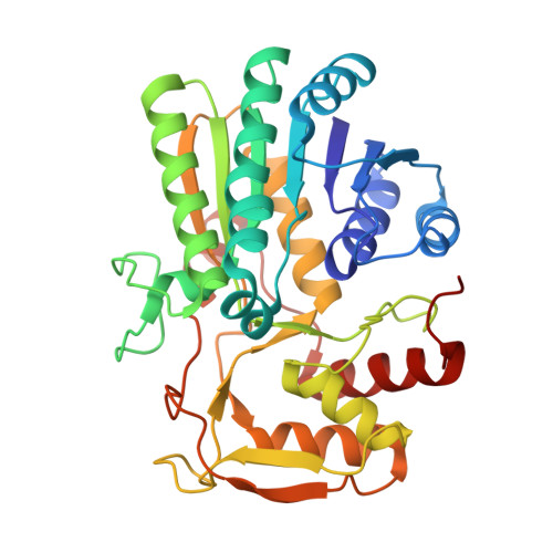
Structural analysis of the Y299C mutant of Escherichia coli UDP-galactose 4-epimerase. Teaching an old dog new tricks.

Thoden, J.B.Henderson, J.M.Fridovich-Keil, J.L.Holden, H.M.

(2002) J Biol Chem 277: 27528-27534

  • DOI: https://doi.org/10.1074/jbc.M204413200
  • Primary Citation of Related Structures:  
    1LRJ, 1LRK, 1LRL

  • PubMed Abstract: 

    UDP-galactose 4-epimerase catalyzes the interconversion of UDP-Gal and UDP-Glc during normal galactose metabolism. The mammalian form of the enzyme, unlike its Escherichia coli counterpart, can also interconvert UDP-GalNAc and UDP-GlcNAc. One key feature of the epimerase reaction mechanism is the rotation of a 4-ketopyranose intermediate in the active site. By comparing the high resolution x-ray structures of both the bacterial and human forms of the enzyme, it was previously postulated that the additional activity in the human epimerase was due to replacement of the structural equivalent of Tyr-299 in the E. coli enzyme with a cysteine residue, thereby leading to a larger active site volume. To test this hypothesis, the Y299C mutant form of the E. coli enzyme was prepared and its three-dimensional structure solved as described here. Additionally, the Y299C mutant protein was assayed for activity against both UDP-Gal and UDP-GalNAc. These studies have revealed that, indeed, this simple mutation did confer UDP-GalNAc/UDP-GlcNAc converting activity to the bacterial enzyme with minimal changes in its three-dimensional structure. Specifically, although the Y299C mutation in the bacterial enzyme resulted in a loss of epimerase activity with regard to UDP-Gal by almost 5-fold, it resulted in a gain of activity against UDP-GalNAc by more than 230-fold.


  • Organizational Affiliation

    Department of Biochemistry, University of Wisconsin, Madison, WI 53706-1544, USA.


Macromolecules
Find similar proteins by:  (by identity cutoff)  |  3D Structure
Entity ID: 1
MoleculeChains Sequence LengthOrganismDetailsImage
UDP-glucose 4-epimerase338Escherichia coliMutation(s): 1 
Gene Names: GALE
EC: 5.1.3.2
UniProt
Find proteins for P09147 (Escherichia coli (strain K12))
Explore P09147 
Go to UniProtKB:  P09147
Entity Groups  
Sequence Clusters30% Identity50% Identity70% Identity90% Identity95% Identity100% Identity
UniProt GroupP09147
Sequence Annotations
Expand
  • Reference Sequence
Experimental Data & Validation

Experimental Data

  • Method: X-RAY DIFFRACTION
  • Resolution: 1.75 Å
  • R-Value Free: 0.247 
  • R-Value Work: 0.171 
  • R-Value Observed: 0.174 
  • Space Group: P 32 2 1
Unit Cell:
Length ( Å )Angle ( ˚ )
a = 83.8α = 90
b = 83.8β = 90
c = 109.6γ = 120
Software Package:
Software NamePurpose
AMoREphasing
TNTrefinement
FRAMBOdata collection
SAINTdata scaling

Structure Validation

View Full Validation Report


Ligand Structure Quality Assessment 


Entry History 

Deposition Data

Revision History  (Full details and data files)

  • Version 1.0: 2002-07-26
    Type: Initial release
  • Version 1.1: 2008-04-28
    Changes: Version format compliance
  • Version 1.2: 2011-07-13
    Changes: Derived calculations, Version format compliance
  • Version 1.3: 2011-11-16
    Changes: Atomic model
  • Version 1.4: 2017-10-11
    Changes: Refinement description
  • Version 2.0: 2021-10-27
    Changes: Atomic model, Database references, Derived calculations
  • Version 2.1: 2024-02-14
    Changes: Advisory, Data collection, Derived calculations, Refinement description