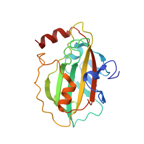
1KN3

Murine PEBP-2 (phosphatidylethanolamine-binding protein-2)


Experimental Data Snapshot

  • Method: X-RAY DIFFRACTION
  • Resolution: 1.80 Å
  • R-Value Free: 0.267 
  • R-Value Work: 0.210 
  • R-Value Observed: 0.213 

wwPDB Validation   3D Report Full Report


This is version 1.3 of the entry. See complete history


Literature

The crystal structure of PEBP-2, a homologue of the PEBP/RKIP family.

Simister, P.C.Banfield, M.J.Brady, R.L.

(2002) Acta Crystallogr D Biol Crystallogr 58: 1077-1080

  • DOI: https://doi.org/10.1107/s090744490200522x
  • Primary Citation of Related Structures:  
    1KN3

  • PubMed Abstract: 

    Proteins from the PEBP (phosphatidylethanolamine-binding protein) family have been identified in a wide variety of species and are thought to regulate a range of intracellular signalling cascades. The rat homologue (known as RKIP; Raf-1 kinase inhibitor protein) has been shown to negatively regulate the MAP kinase pathway through formation of inhibitory complexes with Raf-1 and MEK. The crystal structure of a new, murine member of the PEBP family, termed mPEBP-2, has been determined. On the basis of amino-acid homology, mPEBP-2 belongs to a distinct subset of the mammalian PEBP proteins. Nonetheless, mPEBP-2 is seen to be very similar in structure to other PEBP proteins from human, bovine and plant sources. Regions of distinctive sequence associated with the PEBP-2 subset are discussed with reference to this structure.


  • Organizational Affiliation

    Department of Biochemistry, University of Bristol, Bristol BS8 1TD, England.


Macromolecules
Find similar proteins by:  (by identity cutoff)  |  3D Structure
Entity ID: 1
MoleculeChains Sequence LengthOrganismDetailsImage
Phosphatidylethanolamine Binding Protein-2183Mus musculusMutation(s): 0 
Gene Names: pebp-2
UniProt
Find proteins for Q8VIN1 (Mus musculus)
Explore Q8VIN1 
Go to UniProtKB:  Q8VIN1
Entity Groups  
Sequence Clusters30% Identity50% Identity70% Identity90% Identity95% Identity100% Identity
UniProt GroupQ8VIN1
Sequence Annotations
Expand
  • Reference Sequence
Experimental Data & Validation

Experimental Data

  • Method: X-RAY DIFFRACTION
  • Resolution: 1.80 Å
  • R-Value Free: 0.267 
  • R-Value Work: 0.210 
  • R-Value Observed: 0.213 
  • Space Group: P 21 21 21
Unit Cell:
Length ( Å )Angle ( ˚ )
a = 40.816α = 90
b = 51.401β = 90
c = 79.765γ = 90
Software Package:
Software NamePurpose
AMoREphasing
REFMACrefinement
DENZOdata reduction
SCALEPACKdata scaling

Structure Validation

View Full Validation Report


Entry History 

Deposition Data

Revision History  (Full details and data files)

  • Version 1.0: 2002-06-12
    Type: Initial release
  • Version 1.1: 2008-04-27
    Changes: Version format compliance
  • Version 1.2: 2011-07-13
    Changes: Advisory, Version format compliance
  • Version 1.3: 2023-08-16
    Changes: Data collection, Database references, Refinement description