1KMB

SELECTIN-LIKE MUTANT OF MANNOSE-BINDING PROTEIN A


Experimental Data Snapshot

  • Method: X-RAY DIFFRACTION
  • Resolution: 2.10 Å
  • R-Value Free: 0.265 
  • R-Value Work: 0.202 
  • R-Value Observed: 0.202 

wwPDB Validation   3D Report Full Report


This is version 1.4 of the entry. See complete history


Literature

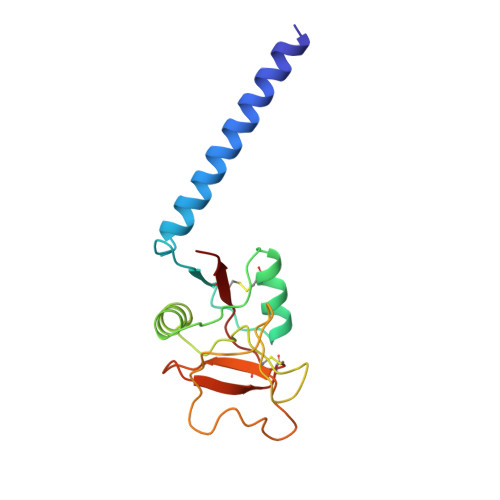
Structure of a selectin-like mutant of mannose-binding protein complexed with sialylated and sulfated Lewis(x) oligosaccharides.

Ng, K.K.Weis, W.I.

(1997) Biochemistry 36: 979-988

  • DOI: https://doi.org/10.1021/bi962564e
  • Primary Citation of Related Structures:  
    1KMB, 2KMB, 3KMB, 4KMB

  • PubMed Abstract: 

    Rat serum mannose-binding protein in which residues 211-213 have been changed to the Lys-Lys-Lys sequence found in E-selectin binds HL-60 cells and the oligosaccharide 3'-NeuAc-Le(x). To understand how this mutant, designated K3, mimics the carbohydrate-binding properties of E-selectin, structures of K3 alone and in complexes with 3'-NeuAc-Le(x), 3'-sulfo-Le(x) and 4'-sulfo-Le(x) have been determined at 1.95-2.1 A resolution by X-ray crystallography. The region of K3 that interacts with bound oligosaccharides superimposes closely with the corresponding region of unliganded E-selectin. In each of the oligosaccharide-protein complexes, the 2- and 3-OH of Fuc coordinate Ca2+ and form a network of cooperative hydrogen bonds with amino acid side chains that also coordinate the Ca2+. Lys211 of the K3 mutant, which corresponds to Lys111 of E-selectin, interacts with each of the three bound ligands: the N zeta atom donates a hydrogen bond to the 4-OH of Gal in 3'-NeuAc-Le(x), forms a water-mediated hydrogen bond with the 4-OH of Gal in 3'-sulfo-Le(x), and forms a salt bridge with the sulfate group of 4'-sulfo-Le(x). Lys213 packs against an otherwise exposed aromatic residue and forms a water-mediated hydrogen bond with Lys211 which may help to position that residue for interactions with bound oligosaccharides. These structures are consistent with previous mutagenesis and chemical modification studies which demonstrate the importance of the Ca2+ ligands as well as Lys111 and Lys113 for carbohydrate binding in the selectins, and they provide a structural basis for understanding the selective recognition of negatively charged Le(x) derivatives by the selectins.


  • Organizational Affiliation

    Department of Structural Biology, Stanford University School of Medicine, California 94305, USA.


Macromolecules
Find similar proteins by:  (by identity cutoff)  |  3D Structure
Entity ID: 1
MoleculeChains Sequence LengthOrganismDetailsImage
MANNOSE-BINDING PROTEIN-AA [auth 1],
B [auth 2],
C [auth 3]
149Rattus norvegicusMutation(s): 3 
UniProt
Find proteins for P19999 (Rattus norvegicus)
Explore P19999 
Go to UniProtKB:  P19999
Entity Groups  
Sequence Clusters30% Identity50% Identity70% Identity90% Identity95% Identity100% Identity
UniProt GroupP19999
Sequence Annotations
Expand
  • Reference Sequence
Small Molecules
Ligands 2 Unique
IDChains Name / Formula / InChI Key2D Diagram3D Interactions
CA
Query on CA

Download Ideal Coordinates CCD File 
D [auth 1]
E [auth 1]
F [auth 1]
H [auth 2]
I [auth 2]
D [auth 1],
E [auth 1],
F [auth 1],
H [auth 2],
I [auth 2],
J [auth 2],
L [auth 3],
M [auth 3],
N [auth 3]
CALCIUM ION
Ca
BHPQYMZQTOCNFJ-UHFFFAOYSA-N
CL
Query on CL

Download Ideal Coordinates CCD File 
G [auth 1],
K [auth 2],
O [auth 3]
CHLORIDE ION
Cl
VEXZGXHMUGYJMC-UHFFFAOYSA-M
Experimental Data & Validation

Experimental Data

  • Method: X-RAY DIFFRACTION
  • Resolution: 2.10 Å
  • R-Value Free: 0.265 
  • R-Value Work: 0.202 
  • R-Value Observed: 0.202 
  • Space Group: C 1 2 1
Unit Cell:
Length ( Å )Angle ( ˚ )
a = 79.7α = 90
b = 84.9β = 105.3
c = 98.7γ = 90
Software Package:
Software NamePurpose
DENZOdata reduction
SCALEPACKdata scaling
X-PLORmodel building
X-PLORrefinement
X-PLORphasing

Structure Validation

View Full Validation Report


Entry History 

Deposition Data

Revision History  (Full details and data files)

  • Version 1.0: 1997-02-12
    Type: Initial release
  • Version 1.1: 2008-03-24
    Changes: Version format compliance
  • Version 1.2: 2011-07-13
    Changes: Version format compliance
  • Version 1.3: 2021-11-03
    Changes: Database references, Derived calculations
  • Version 1.4: 2023-08-09
    Changes: Refinement description