1KDM

THE CRYSTAL STRUCTURE OF THE HUMAN SEX HORMONE-BINDING GLOBULIN (TETRAGONAL CRYSTAL FORM)


Experimental Data Snapshot

  • Method: X-RAY DIFFRACTION
  • Resolution: 2.35 Å
  • R-Value Free: 0.264 
  • R-Value Work: 0.193 

wwPDB Validation   3D Report Full Report


Ligand Structure Quality Assessment 


This is version 1.2 of the entry. See complete history


Literature

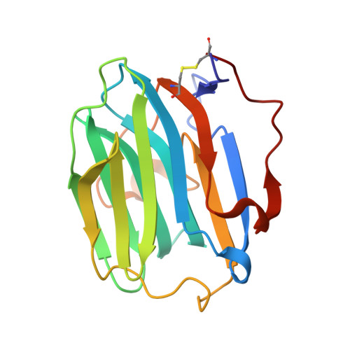
Resolution of a disordered region at the entrance of the human sex hormone-binding globulin steroid-binding site.

Grishkovskaya, I.Avvakumov, G.V.Hammond, G.L.Muller, Y.A.

(2002) J Mol Biol 318: 621-626

  • DOI: https://doi.org/10.1016/S0022-2836(02)00169-9
  • Primary Citation of Related Structures:  
    1KDK, 1KDM

  • PubMed Abstract: 

    The crystal structure of human sex hormone-binding globulin (SHBG) has revealed how 5alpha-dihydrotestosterone intercalates between the two seven-stranded beta-sheets of its amino-terminal laminin G-like domain. However, a region of disorder (residues 130 to 135 of SHBG) was identified together with a zinc-binding site in immediate proximity to the steroid. It has been important to resolve the structure of this region because previous studies have suggested that these residues may contribute to steroid binding directly. Here, we present the 2.35 A and 1.7 A crystal structures of the amino-terminal LG domain of SHBG obtained from a tetragonal crystal form and by EDTA-soaking of a trigonal crystal form, respectively. In both of these new structures, residues Pro130 to Arg135 are now clearly visible. Substitution of the two residues (Leu131Gly and Lys134Ala) pointing towards the steroid has shown that only Leu131 contributes significantly to steroid binding. Rather than covering the steroid-binding pocket in an extended conformation, a 3(10) helical turn is formed by residues Leu131 to Lys134 in this segment. Unfolding of this secondary structure element can either facilitate the entry of the steroids into the binding site or modulate the important contribution that Leu131 makes to steroid binding. A comparison with previous structures supports the concept that zinc binding re-orients the side-chain of His136, and this residue serves as a lever causing disorder within the loop structure between Pro130 and Arg135.


  • Organizational Affiliation

    Forschungsgruppe Kristallographie, Max-Delbrück-Centrum für Molekulare Medizin, 13092 Berlin, Germany.


Macromolecules
Find similar proteins by:  (by identity cutoff)  |  3D Structure
Entity ID: 1
MoleculeChains Sequence LengthOrganismDetailsImage
sex hormone-binding globulin177Homo sapiensMutation(s): 0 
UniProt & NIH Common Fund Data Resources
Find proteins for P04278 (Homo sapiens)
Explore P04278 
Go to UniProtKB:  P04278
PHAROS:  P04278
GTEx:  ENSG00000129214 
Entity Groups  
Sequence Clusters30% Identity50% Identity70% Identity90% Identity95% Identity100% Identity
UniProt GroupP04278
Sequence Annotations
Expand
  • Reference Sequence
Small Molecules
Ligands 2 Unique
IDChains Name / Formula / InChI Key2D Diagram3D Interactions
DHT
Query on DHT

Download Ideal Coordinates CCD File 
C [auth A]5-ALPHA-DIHYDROTESTOSTERONE
C19 H30 O2
NVKAWKQGWWIWPM-ABEVXSGRSA-N
CA
Query on CA

Download Ideal Coordinates CCD File 
B [auth A]CALCIUM ION
Ca
BHPQYMZQTOCNFJ-UHFFFAOYSA-N
Binding Affinity Annotations 
IDSourceBinding Affinity
DHT BindingDB:  1KDM Kd: 0.18 (nM) from 1 assay(s)
IC50: 3.8 (nM) from 1 assay(s)
Experimental Data & Validation

Experimental Data

  • Method: X-RAY DIFFRACTION
  • Resolution: 2.35 Å
  • R-Value Free: 0.264 
  • R-Value Work: 0.193 
  • Space Group: P 43 2 2
Unit Cell:
Length ( Å )Angle ( ˚ )
a = 52.24α = 90
b = 52.24β = 90
c = 148.53γ = 90
Software Package:
Software NamePurpose
MARdata collection
XDSdata reduction
REFMACrefinement
MARdata reduction
XDSdata scaling

Structure Validation

View Full Validation Report


Ligand Structure Quality Assessment 


Entry History 

Deposition Data

Revision History  (Full details and data files)

  • Version 1.0: 2002-05-15
    Type: Initial release
  • Version 1.1: 2008-04-27
    Changes: Version format compliance
  • Version 1.2: 2011-07-13
    Changes: Version format compliance