1K4Q

Human Glutathione Reductase Inactivated by Peroxynitrite


Experimental Data Snapshot

  • Method: X-RAY DIFFRACTION
  • Resolution: 1.90 Å
  • R-Value Free: 0.204 
  • R-Value Work: 0.166 

wwPDB Validation   3D Report Full Report


Ligand Structure Quality Assessment 


This is version 1.2 of the entry. See complete history


Literature

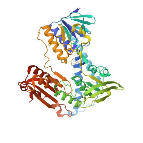
Crystal structure of the antioxidant enzyme glutathione reductase inactivated by peroxynitrite.

Savvides, S.N.Scheiwein, M.Bohme, C.C.Arteel, G.E.Karplus, P.A.Becker, K.Schirmer, R.H.

(2002) J Biol Chem 277: 2779-2784

  • DOI: https://doi.org/10.1074/jbc.M108190200
  • Primary Citation of Related Structures:  
    1K4Q

  • PubMed Abstract: 

    As part of our studies on the nitric oxide-related pathology of cerebral malaria, we show that the antioxidative enzyme glutathione reductase (GR) is inactivated by peroxynitrite, with GR from the malarial parasite Plasmodium falciparum being more sensitive than human GR. The crystal structure of modified human GR at 1.9-A resolution provides the first picture of protein inactivation by peroxynitrite and reveals that this is due to the exclusive nitration of 2 Tyr residues (residues 106 and 114) at the glutathione disulfide-binding site. The selective nitration explains the impairment of binding the peptide substrate and thus the nearly 1000-fold decrease in catalytic efficiency (k(cat)/K(m)) of glutathione reductase observed at physiologic pH. By oxidizing the catalytic dithiol to a disulfide, peroxynitrite itself can act as a substrate of unmodified and bisnitrated P. falciparum glutathione reductase.


  • Organizational Affiliation

    Department of Biochemistry and Biophysics, Oregon State University, Corvallis, Oregon 97331-7305, USA.


Macromolecules
Find similar proteins by:  (by identity cutoff)  |  3D Structure
Entity ID: 1
MoleculeChains Sequence LengthOrganismDetailsImage
Glutathione Reductase461Homo sapiensMutation(s): 2 
EC: 1.6.4.2
UniProt & NIH Common Fund Data Resources
Find proteins for P00390 (Homo sapiens)
Explore P00390 
Go to UniProtKB:  P00390
PHAROS:  P00390
GTEx:  ENSG00000104687 
Entity Groups  
Sequence Clusters30% Identity50% Identity70% Identity90% Identity95% Identity100% Identity
UniProt GroupP00390
Sequence Annotations
Expand
  • Reference Sequence
Small Molecules
Ligands 1 Unique
IDChains Name / Formula / InChI Key2D Diagram3D Interactions
FAD
Query on FAD

Download Ideal Coordinates CCD File 
B [auth A]FLAVIN-ADENINE DINUCLEOTIDE
C27 H33 N9 O15 P2
VWWQXMAJTJZDQX-UYBVJOGSSA-N
Modified Residues  1 Unique
IDChains TypeFormula2D DiagramParent
NIY
Query on NIY
A
L-PEPTIDE LINKINGC9 H10 N2 O5TYR
Experimental Data & Validation

Experimental Data

  • Method: X-RAY DIFFRACTION
  • Resolution: 1.90 Å
  • R-Value Free: 0.204 
  • R-Value Work: 0.166 
  • Space Group: C 1 2 1
Unit Cell:
Length ( Å )Angle ( ˚ )
a = 119.75α = 90
b = 63.65β = 58.42
c = 84.71γ = 90
Software Package:
Software NamePurpose
R-AXISdata collection
SCALEPACKdata scaling
X-PLORmodel building
X-PLORrefinement
R-AXISdata reduction
X-PLORphasing

Structure Validation

View Full Validation Report


Ligand Structure Quality Assessment 


Entry History 

Deposition Data

Revision History  (Full details and data files)

  • Version 1.0: 2002-01-30
    Type: Initial release
  • Version 1.1: 2007-10-16
    Changes: Version format compliance
  • Version 1.2: 2011-07-13
    Changes: Atomic model, Derived calculations, Version format compliance