1JVK

THREE-DIMENSIONAL STRUCTURE OF AN IMMUNOGLOBULIN LIGHT CHAIN DIMER ACTING AS A LETHAL AMYLOID PRECURSOR


Experimental Data Snapshot

  • Method: X-RAY DIFFRACTION
  • Resolution: 1.94 Å
  • R-Value Free: 0.254 
  • R-Value Work: 0.221 
  • R-Value Observed: 0.223 

wwPDB Validation   3D Report Full Report


This is version 2.0 of the entry. See complete history


Literature

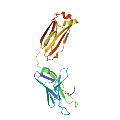
Three-dimensional structure of an immunoglobulin light-chain dimer with amyloidogenic properties.

Bourne, P.C.Ramsland, P.A.Shan, L.Fan, Z.C.DeWitt, C.R.Shultz, B.B.Terzyan, S.S.Moomaw, C.R.Slaughter, C.A.Guddat, L.W.Edmundson, A.B.

(2002) Acta Crystallogr D Biol Crystallogr 58: 815-823

  • DOI: https://doi.org/10.1107/s0907444902004183
  • Primary Citation of Related Structures:  
    1JVK

  • PubMed Abstract: 

    The X-ray structure of an immunoglobulin light-chain dimer isolated from the urine as a "Bence-Jones protein" from a patient with multiple myeloma and amyloidosis (Sea) was determined at 1.94 A resolution and refined to R and R(free) factors of 0.22 and 0.25, respectively. This "amyloidogenic" protein crystallized in the orthorhombic P2(1)2(1)2(1) space group with unit-cell parameters a = 48.28, b = 83.32, c = 112.59 A as determined at 100 K. In the vital organs (heart and kidneys), the equivalent of the urinary protein produced fibrillar amyloid deposits which were fatal to the patient. Compared with the amyloidogenic Mcg light-chain dimer, the Sea protein was highly soluble in aqueous solutions and only crystallized at concentrations approaching 100 mg ml(-1). Both the Sea and Mcg proteins packed into crystals in highly ordered arrangements typical of strongly diffracting crystals of immunoglobulin fragments. Overall similarities and significant differences in the three-dimensional structures and crystalline properties are discussed for the Sea and Mcg Bence-Jones proteins, which together provide a generalized model of abnormalities present in lambda chains, facilitating a better understanding of amyloidosis of light-chain origin (AL).


  • Organizational Affiliation

    OMRF, 825 N E 13th Street, Oklahoma City, OK 73104, USA.


Macromolecules
Find similar proteins by:  (by identity cutoff)  |  3D Structure
Entity ID: 1
MoleculeChains Sequence LengthOrganismDetailsImage
IMMUNOGLOBULIN LAMBDA LIGHT CHAIN
A, B
216Homo sapiensMutation(s): 0 
UniProt
Find proteins for Q6PJG0 (Homo sapiens)
Explore Q6PJG0 
Go to UniProtKB:  Q6PJG0
Entity Groups  
Sequence Clusters30% Identity50% Identity70% Identity90% Identity95% Identity100% Identity
UniProt GroupQ6PJG0
Sequence Annotations
Expand
  • Reference Sequence
Small Molecules
Modified Residues  1 Unique
IDChains TypeFormula2D DiagramParent
PCA
Query on PCA
A, B
L-PEPTIDE LINKINGC5 H7 N O3GLN
Experimental Data & Validation

Experimental Data

  • Method: X-RAY DIFFRACTION
  • Resolution: 1.94 Å
  • R-Value Free: 0.254 
  • R-Value Work: 0.221 
  • R-Value Observed: 0.223 
  • Space Group: P 21 21 21
Unit Cell:
Length ( Å )Angle ( ˚ )
a = 48.28α = 90
b = 83.32β = 90
c = 112.591γ = 90
Software Package:
Software NamePurpose
DENZOdata reduction
SCALEPACKdata scaling
AMoREphasing
REFMACrefinement

Structure Validation

View Full Validation Report


Entry History 

Deposition Data

Revision History  (Full details and data files)

  • Version 1.0: 2002-05-03
    Type: Initial release
  • Version 1.1: 2008-04-27
    Changes: Version format compliance
  • Version 1.2: 2011-07-13
    Changes: Version format compliance
  • Version 2.0: 2019-12-25
    Changes: Derived calculations, Polymer sequence