1JUS

Crystal structure of the multidrug binding transcriptional repressor QacR bound to rhodamine 6G


Experimental Data Snapshot

  • Method: X-RAY DIFFRACTION
  • Resolution: 2.84 Å
  • R-Value Free: 0.272 
  • R-Value Work: 0.230 

wwPDB Validation   3D Report Full Report


Ligand Structure Quality Assessment 


This is version 1.3 of the entry. See complete history


Literature

Structural mechanisms of QacR induction and multidrug recognition.

Schumacher, M.A.Miller, M.C.Grkovic, S.Brown, M.H.Skurray, R.A.Brennan, R.G.

(2001) Science 294: 2158-2163

  • DOI: https://doi.org/10.1126/science.1066020
  • Primary Citation of Related Structures:  
    1JT6, 1JTX, 1JTY, 1JUM, 1JUP, 1JUS

  • PubMed Abstract: 

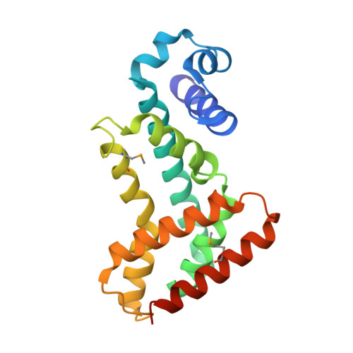
    The Staphylococcus aureus multidrug binding protein QacR represses transcription of the qacA multidrug transporter gene and is induced by structurally diverse cationic lipophilic drugs. Here, we report the crystal structures of six QacR-drug complexes. Compared to the DNA bound structure, drug binding elicits a coil-to-helix transition that causes induction and creates an expansive multidrug-binding pocket, containing four glutamates and multiple aromatic and polar residues. These structures indicate the presence of separate but linked drug-binding sites within a single protein. This multisite drug-binding mechanism is consonant with studies on multidrug resistance transporters.


  • Organizational Affiliation

    Department of Biochemistry and Molecular Biology, Oregon Health & Science University, Portland, OR 97201, USA.


Macromolecules
Find similar proteins by:  (by identity cutoff)  |  3D Structure
Entity ID: 1
MoleculeChains Sequence LengthOrganismDetailsImage
HYPOTHETICAL TRANSCRIPTIONAL REGULATOR IN QACA 5'REGIONA [auth B],
B [auth D],
C [auth A],
D [auth E]
194Staphylococcus aureusMutation(s): 2 
UniProt
Find proteins for P0A0N4 (Staphylococcus aureus)
Explore P0A0N4 
Go to UniProtKB:  P0A0N4
Entity Groups  
Sequence Clusters30% Identity50% Identity70% Identity90% Identity95% Identity100% Identity
UniProt GroupP0A0N4
Sequence Annotations
Expand
  • Reference Sequence
Small Molecules
Ligands 2 Unique
IDChains Name / Formula / InChI Key2D Diagram3D Interactions
RHQ
Query on RHQ

Download Ideal Coordinates CCD File 
W [auth A]RHODAMINE 6G
C28 H31 N2 O3
IWWWBRIIGAXLCJ-KRUMMXJUSA-O
SO4
Query on SO4

Download Ideal Coordinates CCD File 
AA [auth E]
BA [auth E]
E [auth B]
F [auth B]
G [auth B]
AA [auth E],
BA [auth E],
E [auth B],
F [auth B],
G [auth B],
H [auth B],
I [auth B],
J [auth B],
K [auth D],
L [auth D],
M [auth D],
N [auth D],
O [auth D],
P [auth A],
Q [auth A],
R [auth A],
S [auth A],
T [auth A],
U [auth A],
V [auth A],
X [auth E],
Y [auth E],
Z [auth E]
SULFATE ION
O4 S
QAOWNCQODCNURD-UHFFFAOYSA-L
Modified Residues  1 Unique
IDChains TypeFormula2D DiagramParent
MSE
Query on MSE
A [auth B],
B [auth D],
C [auth A],
D [auth E]
L-PEPTIDE LINKINGC5 H11 N O2 SeMET
Experimental Data & Validation

Experimental Data

  • Method: X-RAY DIFFRACTION
  • Resolution: 2.84 Å
  • R-Value Free: 0.272 
  • R-Value Work: 0.230 
  • Space Group: P 42 21 2
Unit Cell:
Length ( Å )Angle ( ˚ )
a = 172.3α = 90
b = 172.3β = 90
c = 95γ = 90
Software Package:
Software NamePurpose
CNSrefinement
MOSFLMdata reduction
CCP4data scaling
CNSphasing

Structure Validation

View Full Validation Report


Ligand Structure Quality Assessment 


Entry History 

Deposition Data

Revision History  (Full details and data files)

  • Version 1.0: 2001-12-12
    Type: Initial release
  • Version 1.1: 2008-04-27
    Changes: Version format compliance
  • Version 1.2: 2011-07-13
    Changes: Version format compliance
  • Version 1.3: 2021-10-27
    Changes: Database references, Derived calculations