1J0R

Crystal structure of the replication termination protein mutant C110S


Experimental Data Snapshot

  • Method: X-RAY DIFFRACTION
  • Resolution: 2.50 Å
  • R-Value Free: 0.279 
  • R-Value Work: 0.233 

wwPDB Validation   3D Report Full Report


This is version 1.4 of the entry. See complete history


Literature

The impact of single cysteine residue mutations on the replication terminator protein

Vivian, J.P.Hastings, A.F.Duggin, I.G.Wake, R.G.Wilce, M.C.J.Wilce, J.A.

(2003) Biochem Biophys Res Commun 310: 1096-1103

  • DOI: https://doi.org/10.1016/j.bbrc.2003.09.126
  • Primary Citation of Related Structures:  
    1J0R

  • PubMed Abstract: 

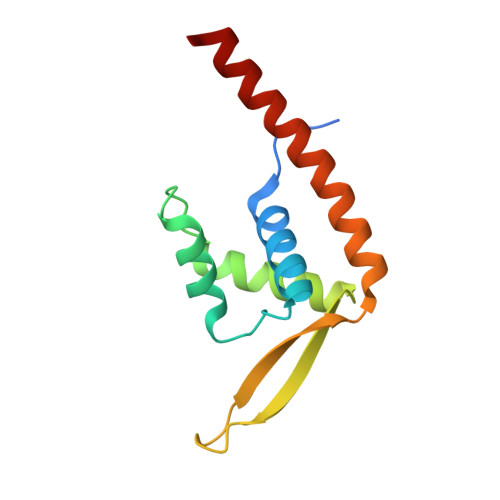
    We report the structural and biophysical consequences of cysteine substitutions in the DNA-binding replication terminator protein (RTP) of Bacillus subtilis, that resulted in an optimised RTP mutant suitable for structural studies. The cysteine residue 110 was replaced with alanine, valine or serine. Protein secondary structure and stability (using circular dichroism spectropolarimetry), self-association (using analytical ultracentrifugation), and DNA-binding measurements revealed RTP.C110S to be the most similar mutant to wild-type RTP. The C110A and C110V.RTP mutants were less soluble, less stable and showed lower DNA-binding affinity. The structure of RTP.C110S, solved to 2.5A resolution using crystallographic methods, showed no major structural perturbation due to the mutation. Heteronuclear NMR spectroscopic studies revealed subtle differences in the electronic environment about the site of mutation. The study demonstrates the suitability of serine as a substitute for cysteine in RTP and the high sensitivity of protein behaviour to single amino acid substitutions.


  • Organizational Affiliation

    Department of Pharmacology, Western Australian Institute for Medical Research, University of Western Australia, Perth, WA 6009, Australia.


Macromolecules
Find similar proteins by:  (by identity cutoff)  |  3D Structure
Entity ID: 1
MoleculeChains Sequence LengthOrganismDetailsImage
replication termination protein
A, B
122Bacillus subtilisMutation(s): 1 
UniProt
Find proteins for P0CI76 (Bacillus subtilis (strain 168))
Explore P0CI76 
Go to UniProtKB:  P0CI76
Entity Groups  
Sequence Clusters30% Identity50% Identity70% Identity90% Identity95% Identity100% Identity
UniProt GroupP0CI76
Sequence Annotations
Expand
  • Reference Sequence
Experimental Data & Validation

Experimental Data

  • Method: X-RAY DIFFRACTION
  • Resolution: 2.50 Å
  • R-Value Free: 0.279 
  • R-Value Work: 0.233 
  • Space Group: C 1 2 1
Unit Cell:
Length ( Å )Angle ( ˚ )
a = 76.88α = 90
b = 50.94β = 91.17
c = 69.65γ = 90
Software Package:
Software NamePurpose
DENZOdata reduction
SCALEPACKdata scaling
CNSrefinement
CNSphasing

Structure Validation

View Full Validation Report


Entry History 

Deposition Data

Revision History  (Full details and data files)

  • Version 1.0: 2003-11-20
    Type: Initial release
  • Version 1.1: 2007-10-16
    Changes: Version format compliance
  • Version 1.2: 2011-07-13
    Changes: Version format compliance
  • Version 1.3: 2021-11-10
    Changes: Database references
  • Version 1.4: 2023-10-25
    Changes: Data collection, Refinement description