The Fab and Fc fragments of IgA1 exhibit a different arrangement from that in IgG: a study by X-ray and neutron solution scattering and homology modelling.
Boehm, M.K., Woof, J.M., Kerr, M.A., Perkins, S.J.(1999) J Mol Biol 286: 1421-1447
- PubMed: 10064707
- DOI: https://doi.org/10.1006/jmbi.1998.2556
- Primary Citation of Related Structures:
1IGA - PubMed Abstract:
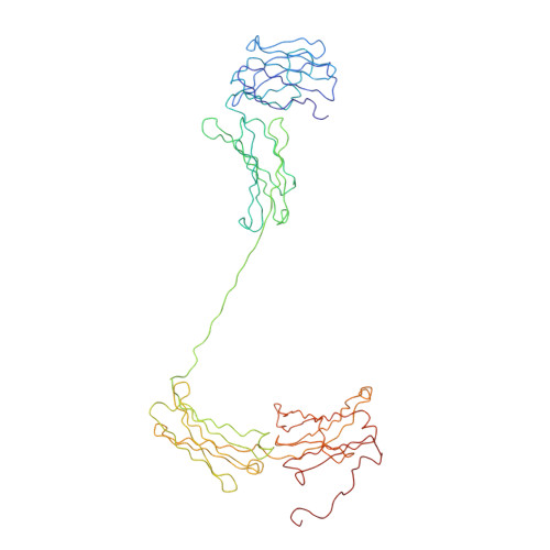
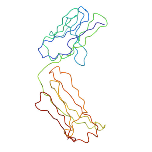
Human immunoglobulin A (IgA) is an abundant antibody that mediates immune protection at mucosal surfaces as well as in plasma. The IgA1 isotype contains two four-domain Fab fragments and a four-domain Fc fragment analogous to that in immunoglobulin G (IgG), linked by a glycosylated hinge region made up of 23 amino acid residues from each of the heavy chains. IgA1 also has two 18 residue tailpieces at the C terminus of each heavy chain in the Fc fragment. X-ray scattering using H2O buffers and neutron scattering using 100 % 2H2O buffers were performed on monomeric IgA1 and a recombinant IgA1 that lacks the tailpiece (PTerm455). The radii of gyration RG from Guinier analyses were similar at 6.11-6.20 nm for IgA1 and 5.84-6.16 nm for PTerm455, and their cross-sectional radii of gyration RXS were also similar. The similarity of the RG and RXS values suggests that the tailpiece of IgA1 is not extended outwards in solution. The IgA1 RG values are higher than those for IgG, and the distance distribution function P(r) showed two distinct peaks, whereas a single peak was observed for IgG. Both results show that the hinge of IgA1 results in an extended Fab and Fc arrangement that is different from that in IgG. Automated curve-fit searches constrained by homology models for the Fab and Fc fragments were used to model the experimental IgA1 scattering curves. A translational search to optimise the relative arrangement of the Fab and Fc fragments held in a fixed orientation resembling that in IgG was not successful in fitting the scattering data. A new molecular dynamics curve-fit search method generated IgA1 hinge structures to which the Fab and Fc fragments could be connected in any orientation. A search based on these identified a limited family of IgA1 structures that gave good curve fits to the experimental data. These contained extended hinges of length about 7 nm that positioned the Fab-to-Fab centre-to-centre separation 17 nm apart while keeping the corresponding Fab-to-Fc separation at 9 nm. The resulting extended T-shaped IgA1 structures are distinct from IgG structures previously determined by scattering and crystallography which have Fab-to-Fab and Fab-to-Fc centre-to-centre separations of 7-9 nm and 6-8 nm, respectively. It was concluded that the IgA1 hinge is structurally distinct from that in IgG, and this results in a markedly different antibody structure that may account for a unique immune role of monomeric IgA1 in plasma and mucosa.
Organizational Affiliation:
Department of Biochemistry and Molecular Biology, Royal Free Campus, University College Medical School, London, UK.