


ATP Binding Independent of Metal Cations in Synapsin II
Esser, L., Palnitkar, M., Deisenhofer, J.To be published.
Experimental Data Snapshot
Entity ID: 1 | |||||
|---|---|---|---|---|---|
| Molecule | Chains | Sequence Length | Organism | Details | Image |
| SYNAPSIN II | 309 | Rattus norvegicus | Mutation(s): 0 | 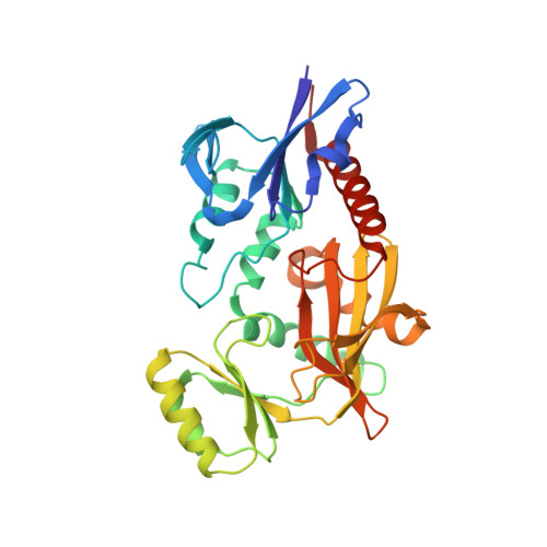 | |
UniProt | |||||
Find proteins for Q63537 (Rattus norvegicus) Explore Q63537 Go to UniProtKB: Q63537 | |||||
Entity Groups | |||||
| Sequence Clusters | 30% Identity50% Identity70% Identity90% Identity95% Identity100% Identity | ||||
| UniProt Group | Q63537 | ||||
Sequence AnnotationsExpand | |||||
| |||||
| Ligands 1 Unique | |||||
|---|---|---|---|---|---|
| ID | Chains | Name / Formula / InChI Key | 2D Diagram | 3D Interactions | |
| ATP Query on ATP | C [auth A], D [auth B] | ADENOSINE-5'-TRIPHOSPHATE C10 H16 N5 O13 P3 ZKHQWZAMYRWXGA-KQYNXXCUSA-N |  | ||
| Length ( Å ) | Angle ( ˚ ) |
|---|---|
| a = 121.275 | α = 90 |
| b = 121.275 | β = 90 |
| c = 165.891 | γ = 120 |
| Software Name | Purpose |
|---|---|
| DENZO | data reduction |
| SCALEPACK | data scaling |
| CNS | refinement |
| CNS | phasing |
RCSB PDB (citation) is hosted by
RCSB PDB is a member of the

