The structure of nucleotidylated histidine-166 of galactose-1-phosphate uridylyltransferase provides insight into phosphoryl group transfer.
Wedekind, J.E., Frey, P.A., Rayment, I.(1996) Biochemistry 35: 11560-11569
- PubMed: 8794735
- DOI: https://doi.org/10.1021/bi9612677
- Primary Citation of Related Structures:
1HXQ - PubMed Abstract:
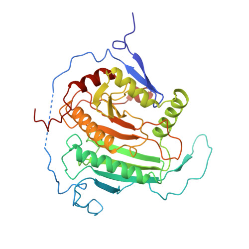
Galactose-1-phosphate uridylyltransferase catalyzes the reaction of UDP-glucose with galactose 1-phosphate to form UDP-galactose and glucose 1-phosphate during normal cellular metabolism. The reaction proceeds through a double displacement mechanism characterized by the formation of a stable nucleotidylated histidine intermediate. This paper describes the preparation of the uridylyl-enzyme complex on the crystalline enzyme from Escherichia coli and its subsequent structure determination by X-ray crystallography. The refined structure has an R-factor of 19.6% (data between 65 and 1.86 A resolution) and reveals modest conformational changes at the active site compared to the inactive UMP/UDP-enzyme complex reported previously [Wedekind, J.E., Frey, P.A., & Rayment, I. (1995) Biochemistry 34, 11049-11061]. In particular, positions of the respective UMP alpha-phosphoryl groups differ by approximately 4 A. Well-defined electron density for the nucleotidylated imidazole supports the existence of a covalent bond between N epsilon 2 of the nucleophile and the alpha-phosphorus of UMP. A hydrogen bond that is conserved in both complexes between His 166 N delta 1 and the carbonyl O of His 164 serves to properly orient the nucleophile and electrostatically stabilize the positively charged imidazolium that results from nucleotidylation. Hydrogen bonds from side-chain Gln 168 to the nonbridging phosphoryl oxygens of the nucleotidyl intermediate appear crucial for the formation and reaction of the uridylyl-enzyme complex as well. The significance of the latter interaction is underscored by the fact that the predominant cause of the metabolic disease galactosemia is the mutation of the corresponding Gln (Gln 188 in humans) to Arg. A comparison to other phosphohistidyl enzymes is described, as well as a revised model for the mechanism of the uridylyltransferase.
Organizational Affiliation:
Institute for Enzyme Research, Graduate School, College of Agricultural and Life Sciences, University of Wisconsin, Madison 53705, USA.