Catalytic Mechanism of Cmp:2-Keto-3-Deoxy-Manno-Octonic Acid Synthetase as Derived from Complexes with Reaction Educt and Product.
Jelakovic, S., Schulz, G.E.(2002) Biochemistry 41: 1174
- PubMed: 11802716
- DOI: https://doi.org/10.1021/bi0119060
- Primary Citation of Related Structures:
1GQ9, 1GQC - PubMed Abstract:
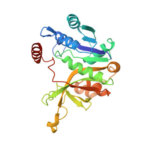
The activation of the sugar 2-keto-3-deoxy-manno-octonic acid (Kdo) is catalyzed by CMP-Kdo synthetase (EC 2.7.7.38) and results in a monophosphate diester with CMP. The enzyme is a pharmaceutical target because CMP-Kdo is required for the biosynthesis of lipopolysaccharides that are vital for Gram-negative bacteria. We have established the structures of an enzyme complex with the educt CTP and of a complex with the product CMP-Kdo by X-ray diffraction analyses at 100 K, both at 2.6 A resolution. The N-terminal domains of the dimeric enzyme bind CTP in a peculiar nucleotide-binding fold with the beta- and gamma-phosphates located at the so-called "PP-loop", whereas the C-terminal domains participate in Kdo binding and in the dimer interface. The unstable nucleotide-sugar CMP-Kdo was produced in a crystal and stabilized by freezing to 100 K. Its formation is accompanied by an induced fit involving mainchain displacements in the 2 A range. The observed binding conformations together with the amino acid conservation pattern during evolution and the putative location of the required Mg(2+) ion suggest a reaction pathway. The enzyme is structurally homologous to the CMP-N-acetylneuraminic acid synthetases in all parts except for the dimer interface. Moreover, the chainfold and the substrate-binding positions resemble those of other enzymes processing nucleotide sugars.
Organizational Affiliation:
Institut für Organische Chemie und Biochemie, Albert-Ludwigs-Universität, Albertstrasse 21, Freiburg im Breisgau, Germany 79104.