1GDV

CRYSTAL STRUCTURE OF CYTOCHROME C6 FROM RED ALGA PORPHYRA YEZOENSIS AT 1.57 A RESOLUTION


Experimental Data Snapshot

  • Method: X-RAY DIFFRACTION
  • Resolution: 1.57 Å
  • R-Value Free: 0.254 
  • R-Value Work: 0.199 

wwPDB Validation   3D Report Full Report


This is version 2.1 of the entry. See complete history


Literature

Structure of cytochrome c6 from the red alga Porphyra yezoensis at 1. 57 A resolution.

Yamada, S.Park, S.Y.Shimizu, H.Koshizuka, Y.Kadokura, K.Satoh, T.Suruga, K.Ogawa, M.Isogai, Y.Nishio, T.Shiro, Y.Oku, T.

(2000) Acta Crystallogr D Biol Crystallogr 56: 1577-1582

  • DOI: https://doi.org/10.1107/s090744490001461x
  • Primary Citation of Related Structures:  
    1GDV

  • PubMed Abstract: 

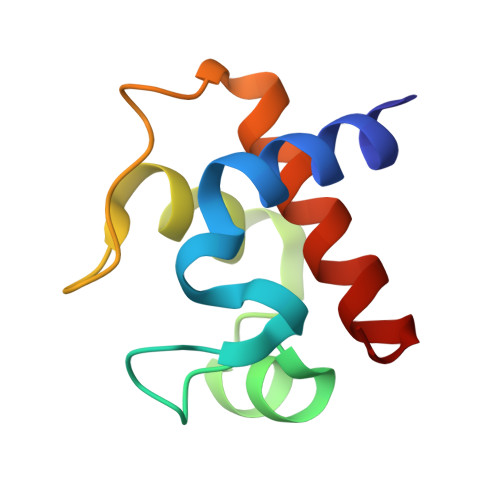
    The crystal structure of cytochrome c(6) from the red alga Porphyra yezoensis has been determined at 1.57 A resolution. The crystal is tetragonal and belongs to space group P4(3)2(1)2, with unit-cell parameters a = b = 49.26 (3), c = 83.45 (4) A and one molecule per asymmetric unit. The structure was solved by the molecular-replacement method and refined with X-PLOR to an R factor of 19.9% and a free R factor of 25.4%. The overall structure of cytochrome c(6) follows the topology of class I c-type cytochromes in which the heme prosthetic group covalently binds to Cys14 and Cys17, and the iron has an octahedral coordination with His18 and Met58 as the axial ligands. The sequence and the structure of the eukaryotic red algal cytochrome c(6) are very similar to those of a prokaryotic cyanobacterial cytochrome c(6) rather than those of eukaryotic green algal c(6) cytochromes.


  • Organizational Affiliation

    Department of Biological Chemistry, College of Bioresource Sciences, Nihon University, 1866 Kameino, Fujisawa, Kanagawa 252-8510, Japan.


Macromolecules
Find similar proteins by:  (by identity cutoff)  |  3D Structure
Entity ID: 1
MoleculeChains Sequence LengthOrganismDetailsImage
CYTOCHROME C685Neopyropia yezoensisMutation(s): 0 
UniProt
Find proteins for Q8WKJ8 (Neopyropia yezoensis)
Explore Q8WKJ8 
Go to UniProtKB:  Q8WKJ8
Entity Groups  
Sequence Clusters30% Identity50% Identity70% Identity90% Identity95% Identity100% Identity
UniProt GroupQ8WKJ8
Sequence Annotations
Expand
  • Reference Sequence
Small Molecules
Ligands 1 Unique
IDChains Name / Formula / InChI Key2D Diagram3D Interactions
HEC
Query on HEC

Download Ideal Coordinates CCD File 
B [auth A]HEME C
C34 H34 Fe N4 O4
HXQIYSLZKNYNMH-LJNAALQVSA-N
Experimental Data & Validation

Experimental Data

  • Method: X-RAY DIFFRACTION
  • Resolution: 1.57 Å
  • R-Value Free: 0.254 
  • R-Value Work: 0.199 
  • Space Group: P 43 21 2
Unit Cell:
Length ( Å )Angle ( ˚ )
a = 49.26α = 90
b = 49.26β = 90
c = 83.45γ = 90
Software Package:
Software NamePurpose
X-PLORmodel building
X-PLORrefinement
MOSFLMdata reduction
CCP4data scaling
X-PLORphasing

Structure Validation

View Full Validation Report


Entry History 

Deposition Data

Revision History  (Full details and data files)

  • Version 1.0: 2001-04-06
    Type: Initial release
  • Version 1.1: 2008-04-27
    Changes: Version format compliance
  • Version 1.2: 2011-07-13
    Changes: Version format compliance
  • Version 2.0: 2019-10-02
    Changes: Atomic model, Data collection, Derived calculations, Non-polymer description, Structure summary
  • Version 2.1: 2023-12-27
    Changes: Data collection, Database references, Derived calculations